Reliability of two different presurgical preparation methods for implant dentistry based on panoramic radiography and cone-beam computed tomography in cadavers
- Affiliations
-
- 1Division in Anatomy and Histology, Department of Oral Biology, Yonsei University College of Dentistry, Seoul, Korea.
- 2Department of Periodontology, Yonsei University College of Dentistry, Seoul, Korea.
- 3Department of Periodontology, Seoul National University School of Dentistry, Seoul, Korea. kst72@snu.ac.kr
- KMID: 2027785
- DOI: http://doi.org/10.5051/jpis.2012.42.2.39
Abstract
- PURPOSE
Special care is necessary to avoid invading important anatomic structures during surgery when presurgical planning is made based on radiographs. However, none of these types of radiography represents a perfect modality. The purpose of this study was to determine the reliability of presurgical planning based on the use of two types of radiographic image (digital panoramic radiography [DPR] and cone-beam computed tomography [CBCT]) by beginner dentists to place implants, and to quantify differences in measurements between radiographic images and real specimens.
METHODS
Ten fresh cadavers without posterior teeth were used, and twelve practitioners who had no experience of implant surgery performed implant surgery after 10 hours of basic instruction using conventional surgical guide based on CBCT or DPR. Two types of measurement error were evaluated: 1) the presurgical measurement error, defined as that between the presurgical and postsurgical measurements in each modality of radiographic analysis, and 2) the measurement error between postsurgical radiography and the real specimen.
RESULTS
The mean presurgical measurement error was significantly smaller for CBCT than for DPR in the maxillary region, whereas it did not differ significantly between the two imaging modalities in the mandibular region. The mean measurement error between radiography and real specimens was significantly smaller for CBCT than for DPR in the maxillary region, but did not differ significantly in the mandibular region.
CONCLUSIONS
Presurgical planning can be performed safely using DPR in the mandible; however, presurgical planning using CBCT is recommended in the maxilla when a structure in a buccolingual location needs to be evaluated because this imaging modality supplies buccolingual information that cannot be obtained from DPR.
MeSH Terms
Figure
-

Figure 1 Measurement of D1. D1 was the difference between the distance from the implant platform to the anatomic structure (e.g., inferior wall of the maxillary sinus or superior border of the mandibular canal) in presurgical cone-beam computed tomography and the distance from the implant platform to the implant apex. M: mandibular canal.

Figure 2 Measurement of D2. D2 was the distance from the implant apex to the anatomic structure in postsurgical cone-beam computed tomography. M: mandibular canal.
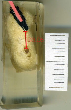
Figure 3 Measurements of D3 and D6. D3 was the distance from the implant apex to the anatomic structure in the real specimen using cone-beam computed tomography as a guide. D6 was same distance in the real specimen using digital panoramic radiography as a guide.
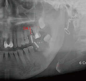
Figure 4 Measurement of D4. D4 was the difference between the distance from the alveolar ridge to the anatomic structure in presurgical digital panoramic radiography and the planned implant length.

Figure 5 Measurement of D5. D5 was the distance from the implant apex to the anatomic structure in postsurgical digital panoramic radiography.
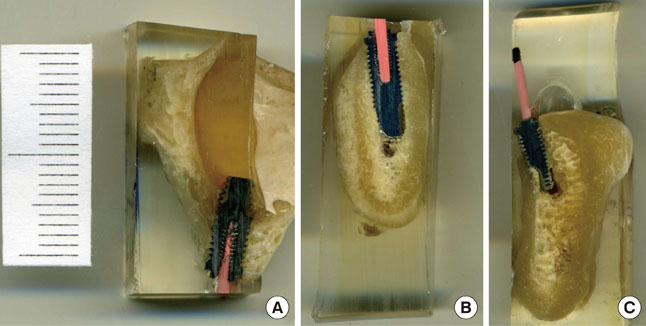
Figure 6 Some critical errors. (A) Maxillary-sinus invasion, (B) mandibular-canal invasion, and (C) lingual-plate perforation.
Cited by 2 articles
-
Commentary on "Reliability of two different presurgical preparation methods for implant dentistry based on panoramic radiography and cone-beam computed tomography in cadavers"
Siamak Sabour
J Periodontal Implant Sci. 2012;42(4):144-144. doi: 10.5051/jpis.2012.42.4.144.Comparing the precision of panoramic radiography and cone-beam computed tomography in avoiding anatomical structures critical to dental implant surgery: A retrospective study
Öznur Özalp, Hüseyin Alican Tezerişener, Burak Kocabalkan, Ulviye Şebnem Büyükkaplan, Mehmet Mustafa Özarslan, Göksel Şimşek Kaya, Mehmet Ali Altay, Alper Sindel
Imaging Sci Dent. 2018;48(4):269-275. doi: 10.5624/isd.2018.48.4.269.
Reference
-
1. Albrektsson T, Dahl E, Enbom L, Engevall S, Engquist B, Eriksson AR, et al. Osseointegrated oral implants: a Swedish multicenter study of 8139 consecutively inserted Nobelpharma implants. J Periodontol. 1988. 59:287–296.2. Misch CE, Perel ML, Wang HL, Sammartino G, Galindo-Moreno P, Trisi P, et al. Implant success, survival, and failure: the International Congress of Oral Implantologists (ICOI) Pisa Consensus Conference. Implant Dent. 2008. 17:5–15.
Article3. Van de Velde T, Glor F, De Bruyn H. A model study on flapless implant placement by clinicians with a different experience level in implant surgery. Clin Oral Implants Res. 2008. 19:66–72.
Article4. Spector L. Computer-aided dental implant planning. Dent Clin North Am. 2008. 52:761–775. vi
Article5. Hong YH, Mun SK. A case of massive maxillary sinus bleeding after dental implant. Int J Oral Maxillofac Surg. 2011. 40:758–760.
Article6. Cho-Lee GY, Naval-Gias L, Castrejon-Castrejon S, Capote-Moreno AL, Gonzalez-Garcia R, Sastre-Perez J, et al. A 12-year retrospective analytic study of the implant survival rate in 177 consecutive maxillary sinus augmentation procedures. Int J Oral Maxillofac Implants. 2010. 25:1019–1027.7. Scaf G, Lurie AG, Mosier KM, Kantor ML, Ramsby GR, Freedman ML. Dosimetry and cost of imaging osseointegrated implants with film-based and computed tomography. Oral Surg Oral Med Oral Pathol Oral Radiol Endod. 1997. 83:41–48.
Article8. Stella JP, Tharanon W. A precise radiographic method to determine the location of the inferior alveolar canal in the posterior edentulous mandible: implications for dental implants. Part 2: Clinical application. Int J Oral Maxillofac Implants. 1990. 5:23–29.9. Reiskin AB. Implant imaging: status, controversies, and new developments. Dent Clin North Am. 1998. 42:47–56.10. Sunden S, Grondahl K, Grondahl HG. Accuracy and precision in the radiographic diagnosis of clinical instability in Branemark dental implants. Clin Oral Implants Res. 1995. 6:220–226.11. Peker I, Alkurt MT, Michcioglu T. The use of 3 different imaging methods for the localization of the mandibular canal in dental implant planning. Int J Oral Maxillofac Implants. 2008. 23:463–470.12. Frei C, Buser D, Dula K. Study on the necessity for cross-section imaging of the posterior mandible for treatment planning of standard cases in implant dentistry. Clin Oral Implants Res. 2004. 15:490–497.
Article13. Tal H, Moses O. A comparison of panoramic radiography with computed tomography in the planning of implant surgery. Dentomaxillofac Radiol. 1991. 20:40–42.
Article14. Bolin A, Eliasson S, von Beetzen M, Jansson L. Radiographic evaluation of mandibular posterior implant sites: correlation between panoramic and tomographic determinations. Clin Oral Implants Res. 1996. 7:354–359.
Article15. Lindh C, Petersson A, Klinge B. Visualisation of the mandibular canal by different radiographic techniques. Clin Oral Implants Res. 1992. 3:90–97.
Article16. Tyndall DA, Brooks SL. Selection criteria for dental implant site imaging: a position paper of the American Academy of Oral and Maxillofacial radiology. Oral Surg Oral Med Oral Pathol Oral Radiol Endod. 2000. 89:630–637.
Article17. Hanazawa T, Sano T, Seki K, Okano T. Radiologic measurements of the mandible: a comparison between CT-reformatted and conventional tomographic images. Clin Oral Implants Res. 2004. 15:226–232.
Article18. Reddy MS, Mayfield-Donahoo T, Vanderven FJ, Jeffcoat MK. A comparison of the diagnostic advantages of panoramic radiography and computed tomography scanning for placement of root form dental implants. Clin Oral Implants Res. 1994. 5:229–238.
Article19. Al-Ekrish AA, Ekram M. A comparative study of the accuracy and reliability of multidetector computed tomography and cone beam computed tomography in the assessment of dental implant site dimensions. Dentomaxillofac Radiol. 2011. 40:67–75.
Article20. Hashimoto K, Arai Y, Iwai K, Araki M, Kawashima S, Terakado M. A comparison of a new limited cone beam computed tomography machine for dental use with a multidetector row helical CT machine. Oral Surg Oral Med Oral Pathol Oral Radiol Endod. 2003. 95:371–377.
Article21. Mozzo P, Procacci C, Tacconi A, Martini PT, Andreis IA. A new volumetric CT machine for dental imaging based on the cone-beam technique: preliminary results. Eur Radiol. 1998. 8:1558–1564.
Article22. Kobayashi K, Shimoda S, Nakagawa Y, Yamamoto A. Accuracy in measurement of distance using limited cone-beam computerized tomography. Int J Oral Maxillofac Implants. 2004. 19:228–231.23. Lindh C, Petersson A, Klinge B. Measurements of distances related to the mandibular canal in radiographs. Clin Oral Implants Res. 1995. 6:96–103.
Article
- Full Text Links
-
- Actions
-
Cited
- CITED
-
- Close
- Share
-
- Similar articles
-
- Commentary on "Reliability of two different presurgical preparation methods for implant dentistry based on panoramic radiography and cone-beam computed tomography in cadavers"
- Reply on "Reliability of two different presurgical preparation methods for implant dentistry based on panoramic radiography and cone-beam computed tomography in cadavers"
- Does cone-beam CT alter treatment plans? Comparison of preoperative implant planning using panoramic versus cone-beam CT images
- Comparing the precision of panoramic radiography and cone-beam computed tomography in avoiding anatomical structures critical to dental implant surgery: A retrospective study
- The value of panoramic radiography in assessing maxillary sinus inflammation
