Does cone-beam CT alter treatment plans? Comparison of preoperative implant planning using panoramic versus cone-beam CT images
- Affiliations
-
- 1OIC, OMFS IMPATH Research Group, Department of Imaging and Pathology, Faculty of Medicine, University of Leuven, Leuven, Belgium. reinhilde.jacobs@uzleuven.be
- 2Department of Oral and Maxillofacial Surgery, University Hospitals, Leuven, Belgium.
- 3Master of Periodontology, Universidad San Martin de Porres, Lima, Peru.
- KMID: 1799593
- DOI: http://doi.org/10.5624/isd.2014.44.2.121
Abstract
- PURPOSE
The present study was performed to compare the planning of implant placement based on panoramic radiography (PAN) and cone-beam computed tomography (CBCT) images, and to study the impact of the image dataset on the treatment planning.
MATERIALS AND METHODS
One hundred five partially edentulous patients (77 males, 28 females, mean age: 46 years, range: 26-67 years) seeking oral implant rehabilitation were referred for presurgical imaging. Imaging consisted of PAN and CBCT imaging. Four observers planned implant treatment based on the two-dimensional (2D) image datasets and at least one month later on the three-dimensional (3D) image dataset. Apart from presurgical diagnostic and dimensional measurement tasks, the observers needed to indicate the surgical confidence levels and assess the image quality in relation to the presurgical needs.
RESULTS
All observers confirmed that both imaging modalities (PAN and CBCT) gave similar values when planning implant diameter. Also, the results showed no differences between both imaging modalities for the length of implants with an anterior location. However, significant differences were found in the length of implants with a posterior location. For implant dimensions, longer lengths of the implants were planned with PAN, as confirmed by two observers. CBCT provided images with improved scores for subjective image quality and surgical confidence levels.
CONCLUSION
Within the limitations of this study, there was a trend toward PAN-based preoperative planning of implant placement leading towards the use of longer implants within the posterior jaw bone.
MeSH Terms
Figure
-
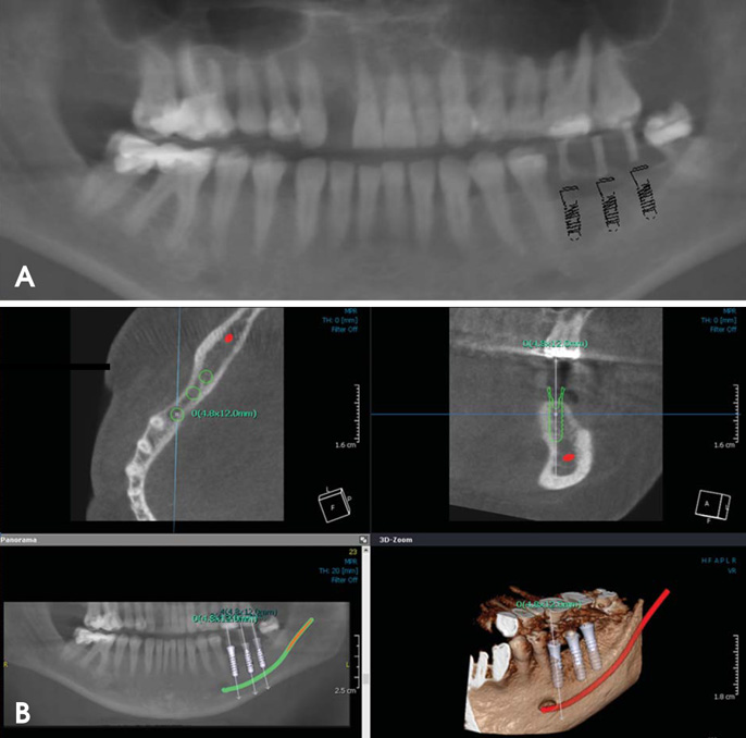
Fig. 1 The implant planning process is performed using panoramic radiography (PAN) (A) and cone-beam computed tomography (CBCT). (B) Images of a 32-year-old woman. After careful evaluation of the 3D data, an appropriate treatment plan is developed, as seen in the cross-sectional images. The bone width is not evident on the PAN image, whereas a possible fenestration can be predicted thanks to the availability of CBCT.
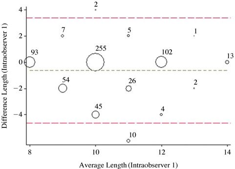
Fig. 2 A graph shows the intra-observer concordance of implant lengths (Bland-Altman).
Cited by 1 articles
-
Preoperative implant planning considering alveolar bone grafting needs and complication prediction using panoramic versus CBCT images
Maria Eugenia Guerrero, Jorge Noriega, Reinhilde Jacobs
Imaging Sci Dent. 2014;44(3):213-220. doi: 10.5624/isd.2014.44.3.213.
Reference
-
1. Belser UC, Schmid B, Higginbottom F, Buser D. Outcome analysis of implant restorations located in the anterior maxilla: a review of the recent literature. Int J Oral Maxillofac Implants. 2004; 19:Suppl. 30–42.2. Lindquist LW, Carlsson GE, Jemt T. A prospective 15-year follow-up study of mandibular fixed prostheses supported by osseointegrated implants. Clinical results and marginal bone loss. Clin Oral Implants Res. 1996; 7:329–336.
Article3. Flanagan D. Important arterial supply of the mandible, control of an arterial hemorrhage, and report of a hemorrhagic incident. J Oral Implantol. 2003; 29:165–173.4. Greenstein G, Cavallaro J, Romanos G, Tarnow D. Clinical recommendations for avoiding and managing surgical complications associated with implant dentistry: a review. J Periodontol. 2008; 79:1317–1329.
Article5. Van Assche N, van Steenberghe D, Guerrero ME, Hirsch E, Schutyser F, Quirynen M, et al. Accuracy of implant placement based on pre-surgical planning of three-dimensional cone-beam images: a pilot study. J Clin Periodontol. 2007; 34:816–821.
Article6. SEDENTEXCT Guideline Development Panel. Radiation protection No 172. Cone beam CT for dental and maxillofacial radiology. Evidence based guidelines. Luxembourg: European Comminssion Directorate-General for Energy;2012.7. Harris D, Horner K, Gröndahl K, Jacobs R, Helmrot E, Benic GI, et al. E.A.O. guidelines for the use of diagnostic imaging in implant dentistry 2011. A consensus workshop organized by the European Association for Osseointegration at the Medical University of Warsaw. Clin Oral Implants Res. 2012; 23:1243–1253.
Article8. Jacobs R, Quirynen M. Dental cone beam CT and its justified use for planning oral implant placement. Periodontol 2000. (in press).9. Bornstein MM, Scarfe WC, Vaughn VM, Jacobs R. Cone beam computed tomography in implant dentistry: a systematic review focusing on guidelines, indications, and radiation dose risks. Int J Oral Maxillofac Implants. 2014; 29:Suppl. 55–77.
Article10. Jacobs R, Adriansens A, Naert I, Quirynen M, Hermans R, Van Steenberghe D. Predictability of reformatted computed tomography for pre-operative planning of endosseous implants. Dentomaxillofac Radiol. 1999; 28:37–41.
Article11. Vazquez L, Nizam Al Din Y, Christoph Belser U, Combescure C, Bernard JP. Reliability of the vertical magnification factor on panoramic radiographs: clinical implications for posterior mandibular implants. Clin Oral Implants Res. 2011; 22:1420–1425.
Article12. Vazquez L, Nizamaldin Y, Combescure C, Nedir R, Bischof M, Dohan Ehrenfest DM, et al. Accuracy of vertical height measurements on direct digital panoramic radiographs using posterior mandibular implants and metal balls as reference objects. Dentomaxillofac Radiol. 2013; 42:20110429.
Article13. Alqerban A, Jacobs R, Fieuws S, Willems G. Comparison of two cone beam computed tomographic systems versus panoramic imaging for localization of impacted maxillary canines and detection of root resorption. Eur J Orthod. 2011; 33:93–102.
Article14. Gerlach NL, Meijer GJ, Maal TJ, Mulder J, Rangel FA, Borstlap WA, et al. Reproducibility of 3 different tracing methods based on cone beam computed tomography in determining the anatomical position of the mandibular canal. J Oral Maxillofac Surg. 2010; 68:811–817.
Article15. Vazquez L, Saulacic N, Belser U, Bernard JP. Efficacy of panoramic radiographs in the preoperative planning of posterior mandibular implants: a prospective clinical study of 1527 consecutively treated patients. Clin Oral Implants Res. 2008; 19:81–85.
Article16. Renton T, Dawood A, Shah A, Searson L, Yilmaz Z. Post-implant neuropathy of the trigeminal nerve. A case series. Br Dent J. 2012; 212:E17.
Article17. Benavides E, Rios HF, Ganz SD, An CH, Resnik R, Reardon GT, et al. Use of cone beam computed tomography in implant dentistry: the International Congress of Oral Implantologists consensus report. Implant Dent. 2012; 21:78–86.18. Alsaadi G, Quirynen M, Michiles K, Teughels W, Komárek A, van Steenberghe D. Impact of local and systemic factors on the incidence of failures up to abutment connection with modified surface oral implants. J Clin Periodontol. 2008; 35:51–57.
Article19. Schropp L, Stavropoulos A, Gotfredsen E, Wenzel A. Comparison of panoramic and conventional cross-sectional tomography for preoperative selection of implant size. Clin Oral Implants Res. 2011; 22:424–429.
Article20. Renouard F, Nisand D. Impact of implant length and diameter on survival rates. Clin Oral Implants Res. 2006; 17:Suppl 2. 35–51.
Article21. Herrmann I, Lekholm U, Holm S, Kultje C. Evaluation of patient and implant characteristics as potential prognostic factors for oral implant failures. Int J Oral Maxillofac Implants. 2005; 20:220–230.22. Weng D, Jacobson Z, Tarnow D, Hürzeler MB, Faehn O, Sanavi F, et al. A prospective multicenter clinical trial of 3i machined-surface implants: results after 6 years of follow-up. Int J Oral Maxillofac Implants. 2003; 18:417–423.23. Lemmerman KJ, Lemmerman NE. Osseointegrated dental implants in private practice: a long-term case series study. J Periodontol. 2005; 76:310–319.
Article24. Romeo E, Lops D, Margutti E, Ghisolfi M, Chiapasco M, Vogel G. Long-term survival and success of oral implants in the treatment of full and partial arches: a 7-year prospective study with the ITI dental implant system. Int J Oral Maxillofac Implants. 2004; 19:247–259.25. Jacobs R, Adriansens A, Verstreken K, Suetens P, van Steenberghe D. Predictability of a three-dimensional planning system for oral implant surgery. Dentomaxillofac Radiol. 1999; 28:105–111.
Article26. Frei C, Buser D, Dula K. Study on the necessity for cross-section imaging of the posterior mandible for treatment planning of standard cases in implant dentistry. Clin Oral Implants Res. 2004; 15:490–497.
Article27. Diniz AF, Mendonça EF, Leles CR, Guilherme AS, Cavalcante MP, Silva MA. Changes in the pre-surgical treatment planning using conventional spiral tomography. Clin Oral Implants Res. 2008; 19:249–253.
Article28. Reddy MS, Mayfield-Donahoo T, Vanderven FJ, Jeffcoat MK. A comparison of the diagnostic advantages of panoramic radiography and computed tomography scanning for placement of root form dental implants. Clin Oral Implants Res. 1994; 5:229–238.
Article29. Liang X, Jacobs R, Hassan B, Li L, Pauwels R, Corpas L, et al. A comparative evaluation of cone beam computed tomography (CBCT) and multi-slice CT (MSCT) Part I. On subjective image quality. Eur J Radiol. 2010; 75:265–269.30. Shelley AM, Brunton P, Horner K. Subjective image quality assessment of cross sectional imaging methods for the symphyseal region of the mandible prior to dental implant placement. J Dent. 2011; 39:764–770.
Article31. Loubele M, Guerrero ME, Jacobs R, Suetens P, van Steenberghe D. A comparison of jaw dimensional and quality assessments of bone characteristics with cone-beam CT, spiral tomography, and multi-slice spiral CT. Int J Oral Maxillofac Implants. 2007; 22:446–454.32. Baciut M, Hedesiu M, Bran S, Jacobs R, Nackaerts O, Baciut G. Pre- and postoperative assessment of sinus grafting procedures using cone-beam computed tomography compared with panoramic radiographs. Clin Oral Implants Res. 2013; 24:512–516.
Article
- Full Text Links
-
- Actions
-
Cited
- CITED
-
- Close
- Share
-
- Similar articles
-
- Cone beam CT findings of retromolar canals: Report of cases and literature review
- Assessment of the relationship between the mandibular third molar and the mandibular canal using panoramic radiograph and cone beam computed tomography
- Commentary on "Reliability of two different presurgical preparation methods for implant dentistry based on panoramic radiography and cone-beam computed tomography in cadavers"
- Three-dimensional imaging modalities in endodontics
- Preoperative implant planning considering alveolar bone grafting needs and complication prediction using panoramic versus CBCT images
