J Korean Neurosurg Soc.
2012 Nov;52(5):466-471. 10.3340/jkns.2012.52.5.466.
Magnetoencephalography Interictal Spike Clustering in Relation with Surgical Outcome of Cortical Dysplasia
- Affiliations
-
- 1MEG Center, Department of Neurosurgery, Seoul National University Hospital, Seoul, Korea. chungc@snu.ac.kr
- 2Department of Neurosurgery, Seoul National University College of Medicine, Seoul, Korea.
- 3Neuroscience Research Institute, Seoul National University Medical Research Center, Seoul, Korea.
- 4Research Center for Sensory Organs, Seoul National University, Seoul, Korea.
- KMID: 2018253
- DOI: http://doi.org/10.3340/jkns.2012.52.5.466
Abstract
OBJECTIVE
The aim of this study was to devise an objective clustering method for magnetoencephalography (MEG) interictal spike sources, and to identify the prognostic value of the new clustering method in adult epilepsy patients with cortical dysplasia (CD).
METHODS
We retrospectively analyzed 25 adult patients with histologically proven CD, who underwent MEG examination and surgical resection for intractable epilepsy. The mean postoperative follow-up period was 3.1 years. A hierarchical clustering method was adopted for MEG interictal spike source clustering. Clustered sources were then tested for their prognostic value toward surgical outcome.
RESULTS
Postoperative seizure outcome was Engel class I in 6 (24%), class II in 3 (12%), class III in 12 (48%), and class IV in 4 (16%) patients. With respect to MEG spike clustering, 12 of 25 (48%) patients showed 1 cluster, 2 (8%) showed 2 or more clusters within the same lobe, 10 (40%) showed 2 or more clusters in a different lobe, and 1 (4%) patient had only scattered spikes with no clustering. Patients who showed focal clustering achieved better surgical outcome than distributed cases (p=0.017).
CONCLUSION
This is the first study that introduces an objective method to classify the distribution of MEG interictal spike sources. By using a hierarchical clustering method, we found that the presence of focal clustered spikes predicts a better postoperative outcome in epilepsy patients with CD.
Keyword
MeSH Terms
Figure
-
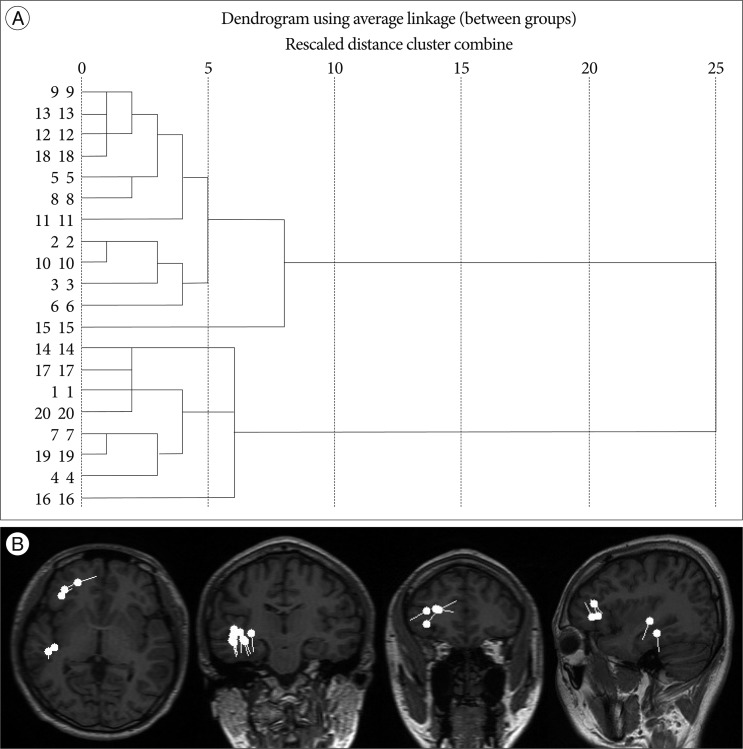
Fig. 1 Hierarchical dendrogram (A) and equivalent current dipoles on MR images (B) for Patient #10. The individual spike sources (n=20) are represented along with the vertical axis. The ⊃-shaped lines connect the spike objects that are combined at each aggregation stage.
Reference
-
1. Berkovic SF, McIntosh AM, Kalnins RM, Jackson GD, Fabinyi GC, Brazenor GA, et al. Preoperative MRI predicts outcome of temporal lobectomy : an actuarial analysis. Neurology. 1995; 45:1358–1363. PMID: 7617198.
Article2. Blümcke I, Thom M, Aronica E, Armstrong DD, Vinters HV, Palmini A, et al. The clinicopathologic spectrum of focal cortical dysplasias : a consensus classification proposed by an ad hoc Task Force of the ILAE Diagnostic Methods Commission. Epilepsia. 2011; 52:158–174. PMID: 21219302.
Article3. Chassoux F, Devaux B, Landré E, Turak B, Nataf F, Varlet P, et al. Stereoelectroencephalography in focal cortical dysplasia : a 3D approach to delineating the dysplastic cortex. Brain. 2000; 123:1733–1751. PMID: 10908202.
Article4. Chung CK, Lee SK, Kim KJ. Surgical outcome of epilepsy caused by cortical dysplasia. Epilepsia. 2005; 46(Suppl 1):25–29. PMID: 15816975.
Article5. Engel JJ. Engel JJ, editor. Outcome with respect to epileptic seizures. Surgical treatment of the epilepsies. 1993. New York: Raven Press;p. 609–621.6. Fischer MJ, Scheler G, Stefan H. Utilization of magnetoencephalography results to obtain favourable outcomes in epilepsy surgery. Brain. 2005; 128:153–157. PMID: 15563514.
Article7. Hirabayashi S, Binnie CD, Janota I, Polkey CE. Surgical treatment of epilepsy due to cortical dysplasia : clinical and EEG findings. J Neurol Neurosurg Psychiatry. 1993; 56:765–770. PMID: 8331351.
Article8. Hudgins RJ, Flamini JR, Palasis S, Cheng R, Burns TG, Gilreath CL. Surgical treatment of epilepsy in children caused by focal cortical dysplasia. Pediatr Neurosurg. 2005; 41:70–76. PMID: 15942276.
Article9. Iida K, Otsubo H, Matsumoto Y, Ochi A, Oishi M, Holowka S, et al. Characterizing magnetic spike sources by using magnetoencephalography-guided neuronavigation in epilepsy surgery in pediatric patients. J Neurosurg. 2005; 102:187–196. PMID: 16156229.
Article10. Kim IM, Son EI. Surgery of intractable epilepsy associated with cortical dysplasia. J Korean Neurosurg Soc. 1999; 28:942–948.11. Kloss S, Pieper T, Pannek H, Holthausen H, Tuxhorn I. Epilepsy surgery in children with focal cortical dysplasia (FCD) : results of long-term seizure outcome. Neuropediatrics. 2002; 33:21–26. PMID: 11930272.
Article12. Kral T, Clusmann H, Blümcke I, Fimmers R, Ostertun B, Kurthen M, et al. Outcome of epilepsy surgery in focal cortical dysplasia. J Neurol Neurosurg Psychiatry. 2003; 74:183–188. PMID: 12531945.
Article13. Lee SK, Lee SY, Kim KK, Hong KS, Lee DS, Chung CK. Surgical outcome and prognostic factors of cryptogenic neocortical epilepsy. Ann Neurol. 2005; 58:525–532. PMID: 16037972.
Article14. Morioka T, Nishio S, Ishibashi H, Muraishi M, Hisada K, Shigeto H, et al. Intrinsic epileptogenicity of focal cortical dysplasia as revealed by magnetoencephalography and electrocorticography. Epilepsy Res. 1999; 33:177–187. PMID: 10094429.
Article15. Oishi M, Kameyama S, Masuda H, Tohyama J, Kanazawa O, Sasagawa M, et al. Single and multiple clusters of magnetoencephalographic dipoles in neocortical epilepsy : significance in characterizing the epileptogenic zone. Epilepsia. 2006; 47:355–364. PMID: 16499760.
Article16. RamachandranNair R, Otsubo H, Shroff MM, Ochi A, Weiss SK, Rutka JT, et al. MEG predicts outcome following surgery for intractable epilepsy in children with normal or nonfocal MRI findings. Epilepsia. 2007; 48:149–157. PMID: 17241222.
Article17. Rosenow F, Lüders H. Presurgical evaluation of epilepsy. Brain. 2001; 124:1683–1700. PMID: 11522572.
Article18. Sisodiya SM. Surgery for malformations of cortical development causing epilepsy. Brain. 2000; 123:1075–1091. PMID: 10825348.
Article19. Spencer SS. Long-term outcome after epilepsy surgery. Epilepsia. 1996; 37:807–813. PMID: 8814092.
Article20. Tassi L, Colombo N, Garbelli R, Francione S, Lo Russo G, Mai R, et al. Focal cortical dysplasia : neuropathological subtypes, EEG, neuroimaging and surgical outcome. Brain. 2002; 125:1719–1732. PMID: 12135964.
Article21. Taulu S, Simola J. Spatiotemporal signal space separation method for rejecting nearby interference in MEG measurements. Phys Med Biol. 2006; 51:1759–1768. PMID: 16552102.
Article22. Taulu S, Simola J, Kajola M. Applications of the signal space separation method. IEEE Trans Signal Process. 2005; 53:3359–3372.
Article23. Tonini C, Beghi E, Berg AT, Bogliun G, Giordano L, Newton RW, et al. Predictors of epilepsy surgery outcome : a meta-analysis. Epilepsy Res. 2004; 62:75–87. PMID: 15519134.24. Widdess-Walsh P, Kellinghaus C, Jeha L, Kotagal P, Prayson R, Bingaman W, et al. Electro-clinical and imaging characteristics of focal cortical dysplasia : correlation with pathological subtypes. Epilepsy Res. 2005; 67:25–33. PMID: 16181772.
Article25. Widjaja E, Otsubo H, Raybaud C, Ochi A, Chan D, Rutka JT, et al. Characteristics of MEG and MRI between Taylor's focal cortical dysplasia (type II) and other cortical dysplasia : surgical outcome after complete resection of MEG spike source and MR lesion in pediatric cortical dysplasia. Epilepsy Res. 2008; 82:147–155. PMID: 18774695.
Article
- Full Text Links
-
- Actions
-
Cited
- CITED
-
- Close
- Share
-
- Similar articles
-
- Magnetoencephalography in Pediatric Lesional Epilepsy Surgery
- MEG and EEG dipole clusters from extended cortical sources
- Magnetoencephalography in Epilepsy
- Cortical Dysplasia and Mesial Temporal Sclerosis in Temporal Lobe Epilepsy Pre-operative Clinical Feature and Surgical Outcome between Patients with Dual Pathology and Patients with Mesial Temporal Sclerosis
- Cortical Dysplasia: Tc-99m ECD SPECT Findings and Comparative Study with MRI according to Pathologic Grading
