A Clinical Study on the Diabetic Foot Wound
- Affiliations
-
- 1Department of Orthopedic Surgery, Eulji Medcal Center, Korea.
- KMID: 2008100
- DOI: http://doi.org/10.4093/jkda.2007.31.1.89
Abstract
-
PURPOSE: To make protocol on diabetic foot ulcer, with making use of this protocol, investigate hospitalized patients who have had diabetic foot wound and define pattern, characteristics and problems of diabetic foot in hospitalized patients
MATERIALS AND METHODS
From Oct. 2002 to Sep. 2003, Seventy-two patients who had been admitted to our hospital due to treatment of diabetic foot wound studied with use of the protocol designed by the authors.
RESULTS
The mean age of patients was 64.3 years and male patients were twice as many as female. The most common cause of hospitalization was infection of diabetic foot (77.7%). As a basic pathology of diabetic foot, the main pathology of diabetic foot was the neuropathy that is four times more than vasculopathy. The causations of wound were infection with no specific cause (40.0%). No statistical difference was found between timing of hospitalization and the results of treatment in vasculopathic group but in neuropathic ulcer group, the major procedure such as amputation, and the times of debridement in operation room and are more common in patients who were hospitalized after 3rd days of beginning of symptoms than within 3rd day.
CONCLUSION
The education that the patient having a foot symptom have to visit the hospital as soon as possible on patients is important to prevent morbidity of diabetic foot wound, long hospitalization and amputation. The protocol that we are presenting can be modified for other study related to diabetic foot.
Keyword
MeSH Terms
Figure
-
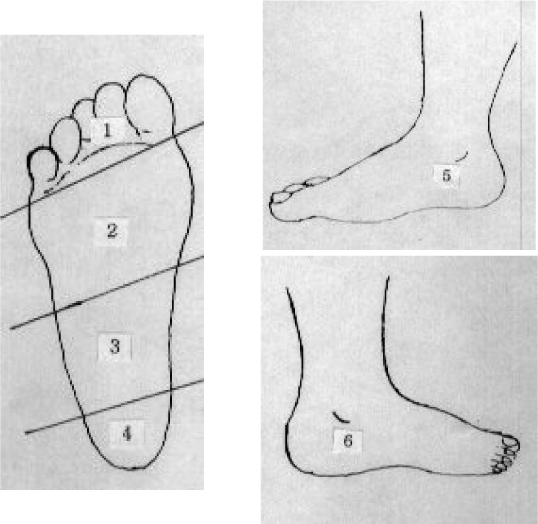
Fig. 1 Location of wound. The location of diabetic foot wound is divided into 6 zone. zone 1, the toe area; zone 2, the metatarsal area; zone 3, the mid-tarsal area; zone 4, the heel; zone 5, the medial malleolar area; zone 6, the lat. malleoular area.
Cited by 3 articles
-
Diabetic Foot
Kwang-Won Kim
J Korean Med Assoc. 2007;50(5):447-454. doi: 10.5124/jkma.2007.50.5.447.The Changes of Trends in the Diagnosis and Treatment of Diabetic Foot Ulcer over a 10-Year Period: Single Center Study
Choong Hee Kim, Jun Sung Moon, Seung Min Chung, Eun Jung Kong, Chul Hyun Park, Woo Sung Yoon, Tae Gon Kim, Woong Kim, Ji Sung Yoon, Kyu Chang Won, Hyoung Woo Lee
Diabetes Metab J. 2018;42(4):308-319. doi: 10.4093/dmj.2017.0076.Factors Contributing to Diabetic Foot Ulcer among Patients with Type 2 Diabetes Mellitus
Seo Jin Park, Taeyoung Yang, Jun Young Lee, Jinhee Kim
Korean J Adult Nurs. 2018;30(1):106-114. doi: 10.7475/KJAN.2018.30.1.106.
Reference
-
1. Zimmet P, Alberti KG, Shaw J. Global and Societal implications of the diabetes epidemic. Nature. 2001. 414:782–787.2. American Diabetic Association. Consensus development conference on diabetic foot wound care. Diabetes Care. 1999. 22:1354–1360.3. Bell-Krotoski J, Tomancik E. The repeatability of testing with Semmes-Weinstein monofilaments. J Hand Surg. 1987. 12:155–161.4. Benotmane A, Mohammedi F, Ayad F, Kadi A, Azzpuz A. Diaetic foot lesions: etiologic and prognostic factors. Diabetes Metab. 2000. 26:113–117.5. Brodsky J. Outpatient diagnosis and care of the diabetic foot. Instr Course Lect. 1993. 42:121–139.6. Frykberg RG, Armstrong DG, Giurini J, Edwards A, Kravette M, Kravitz S, Ross C, Stavosky J, Stuck R, Vanore J. American college of foot and ankle surgeons. Diabetic foot disorders. A clinical practice guideline. J Foot Ankle Surg. 2000. 39:S1–S60.7. Coughlin MJ, Mann RA. Surgery of the foot and ankle: The diabetic foot. 1999. 7th ed. Philadelphia, St. Louis: Mosby;895–969.8. Gale EA, Gillespie KM. Diabetes and gender. Diabetologia. 2001. 44:3–15.9. Smith DG, Barnes BC, Sands AK, Boyko EJ, Ahroni JH. Prevalence of radiographic foot abnormalities in patients with diabetes. Foot Ankle Int. 1997. 18:342–346.10. Rayfield EJ, Ault MJ, Keusch GT, Brothers MJ, Mechemias C, Smith H. Infections and diabetes: the case for glucose control. Am J Med. 1985. 72:439–450.11. Watts SA, Daly B, Anthony M, McDonald P, Khoury A, Dahar W. The effect of age, gender, risk level and glycosylated hemoglobin in predicting foot amputation in HMO patients with diabeties. Am Acad Nurse Pract. 2001. 13:230–235.13. Klenerman L, McCabe C, Cogley D, Crerand S, Laing P, White M. Screening for patients at risk of diabetic foot ulceration in a general diabetic patient clinic. Diabet Med. 1996. 13:561–563.14. Kumar S, Fernando DJ, Veves A, Knowles EA, Young MJ, Boulton AJ. Semmes-Weinstein monofilaments: A simple effective and inexpensive screening device for identifying diabetic patients at risk of foot ulceration. Diabetes Res Clin Pract. 1991. 13(1-2):63–67.15. Shults DW, Hunter GS, McIntyre KE, Parent FN, Piotrowski JJ, Bernhard VM. Value of radiographs and bone scans in determining the need for therapy in diabetic patients with foot ulcers. Am J Surg. 1989. 158:525–529.17. McGill M, Molyneaux L, Yue DK. Use of the Semmes-Weinstein 5.07/10 gram monofilament;the long and the short of it. Diabet Med. 1998. 15(7):615–617.18. Luger E, Nissan M, Karpf A, Steinberg E, Dekel S. Dynamic Pressures on the diabetic foot. Foot Ankle Int. 2001. 22(9):715–719.20. Malone JM, Snyder M, Anderson G, Bernard VM, Holloway GA Jr, Bunt TJ. Prevention of amputation by diabetic education. Am J Surg. 1989. 158(6):520–525.21. Reveal GT, Laughlin RT, Capecci P, Reeve FM. Foot and ankle survey in adults with diabetes mellitus. Foot Ankle Int. 2001. 22(9):739–743.22. Wheat LJ, Allen SD, Henry M, Kernek CB, Siders JA, Kuebler T, Fineberg N, Norton J. Diabetic foot infections; Bacteriologic analysis. Arch Intern Med. 1986. 146:1935–1940.23. Sosenko JM, Gadia MT, Natori N, Ayyar DR, Ramos LB, Skyler JS. Neurofunctional testing for the detection of diabetic peripheral neuropathy. Arch Intern Med. 1987. 147:1741–1744.
- Full Text Links
-
- Actions
-
Cited
- CITED
-
- Close
- Share
-
- Similar articles
-
- Orthotic and Prosthetic Options after Diabetic Foot Amputation
- Latest Approaches in the Treatment of Diabetic Foot Ulcers: An Integrative Analysis Based on the International Working Group on the Diabetic Foot (IWGDF) 2023 Guidelines
- Prevention of Diabetic Foot and Treatment of Pre-Ulcerative Signs
- Diabetic Foot Wounds with Concomitant Skin Malignancies
- Diabetic Foot Education and a Diabetic Foot Screening Tool
