J Korean Bone Joint Tumor Soc.
2010 Jun;16(1):42-46. 10.5292/jkbjts.2010.16.1.42.
Deep Submuscular Parosteal Angiomyxolipoma in a Child
- Affiliations
-
- 1Department of Orthopedic Surgery, Kangnam Sacred Heart Hospital, College of Medicine, Hallym University, Seoul, Korea. dr73@hallym.or.kr
- KMID: 2006341
- DOI: http://doi.org/10.5292/jkbjts.2010.16.1.42
Abstract
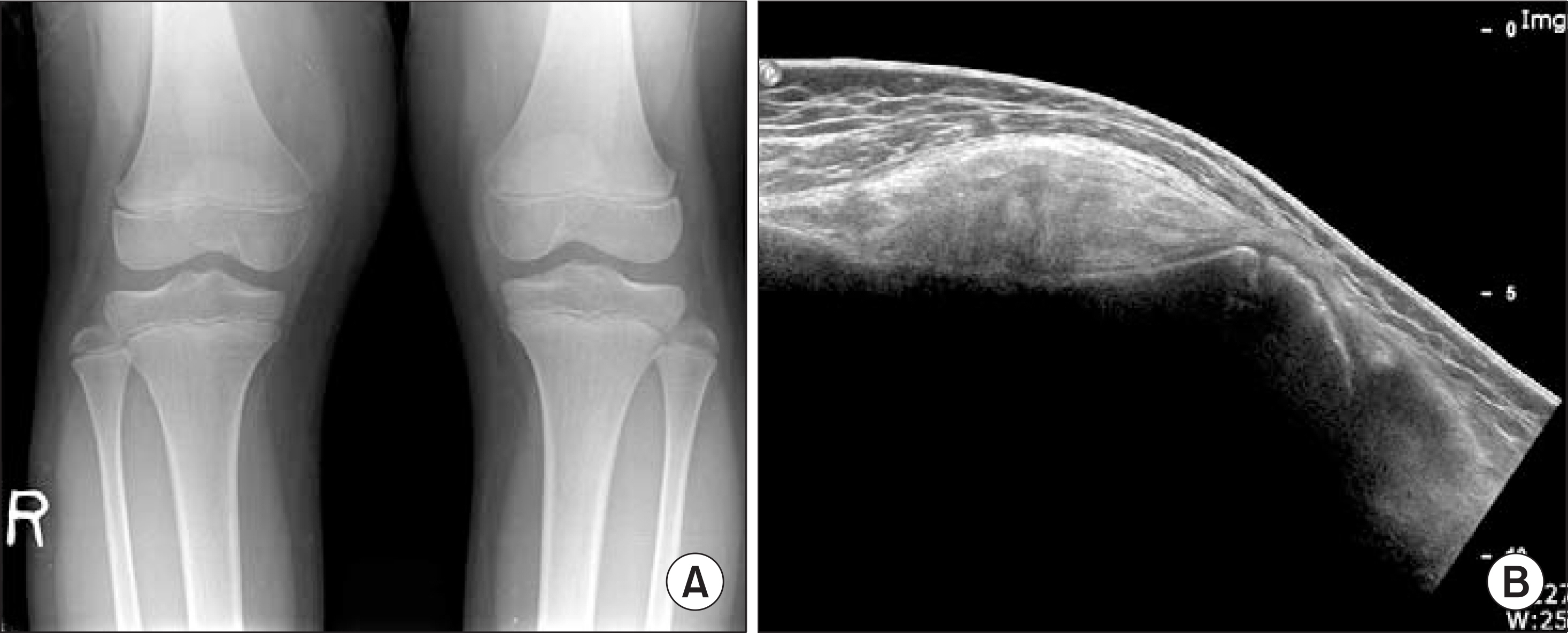
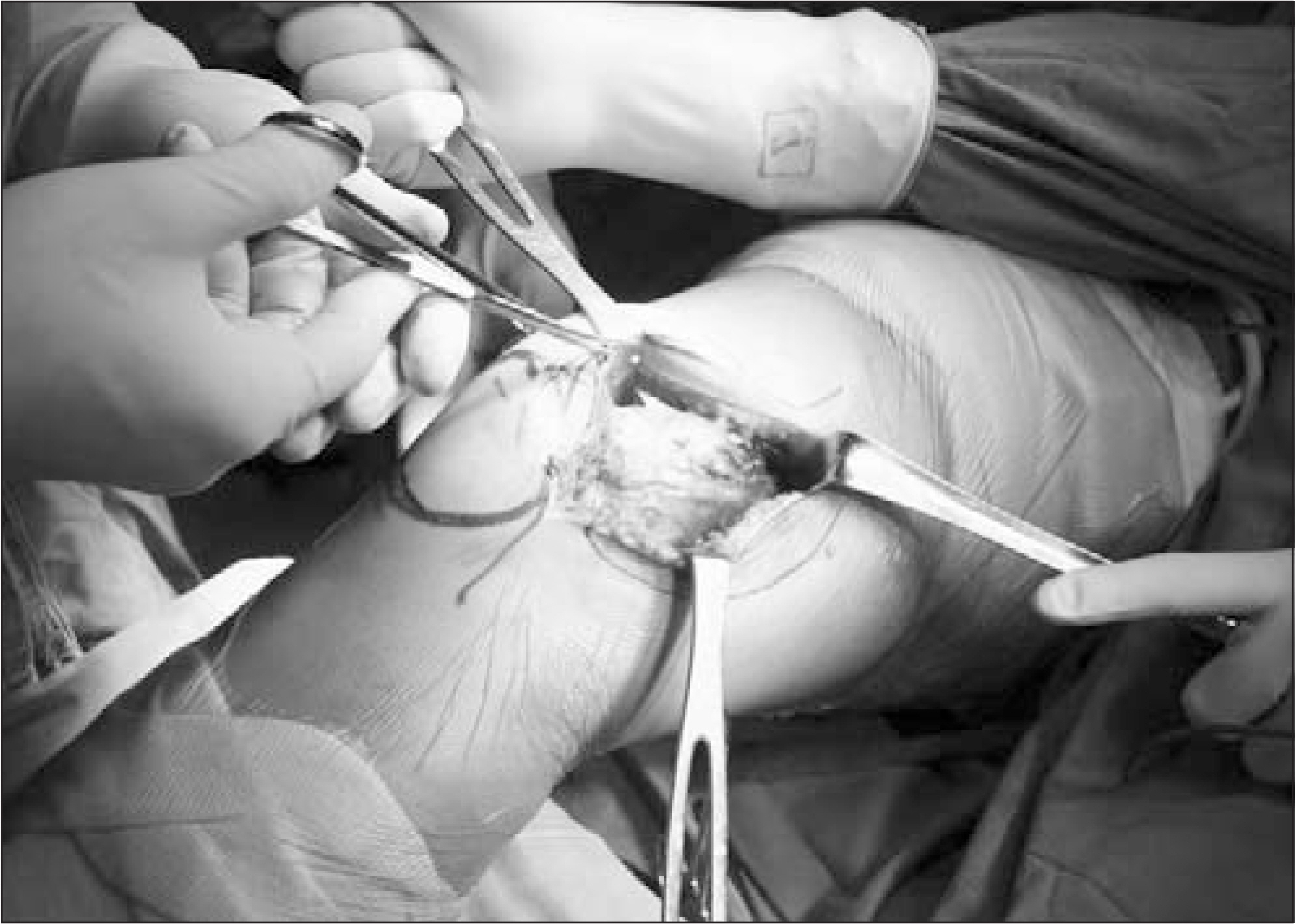
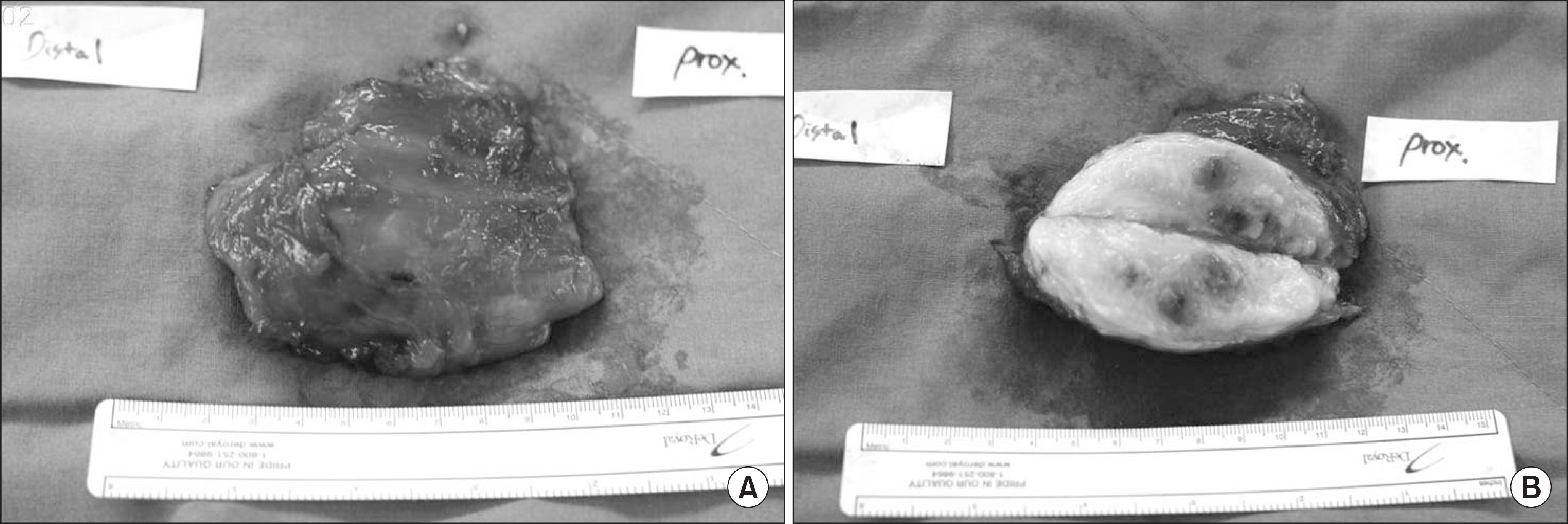
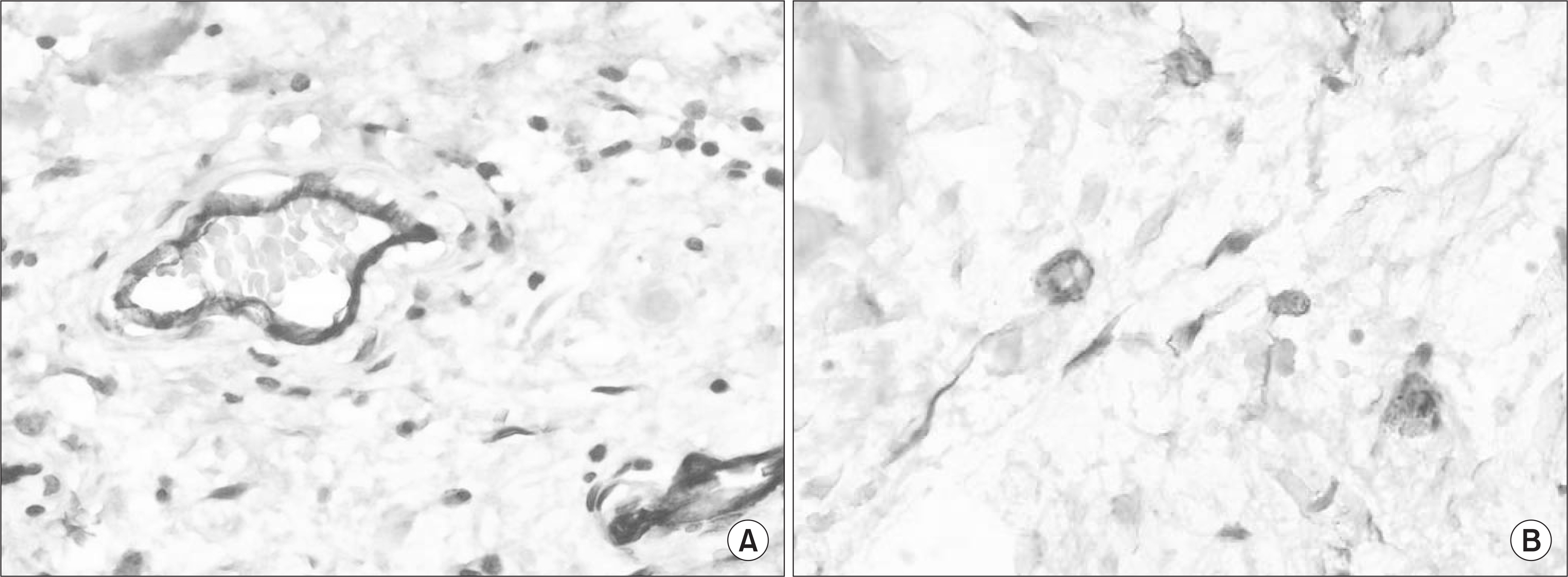
- Angiomyxolipoma is a rare variant of lipoma, which is described by Mai, 1996, at first. The nine cases of which have been reported to date. Microscopically, the lesion consists of adipose tissue with the paucicellular myxoid areas and fat tissue with numerous thin, dilated, and congestive blood vessels. The reported cases mostly located to the superficial layer on the scalp, subungual, extremities in adults. We report one case of angiomyxolipoma located in the submuscular and parosteal area in the distal femur around knee joint in a child.
Keyword
MeSH Terms
Figure
Reference
-
1. Lee HG. Tumor of bone and joint. 1st ed.Seoul: Choisin uehaksa;1996. p. 355–8.2. Mai KT, Yazdi HM, Collins JP. Vascular myxolipoma (“angiomyxolipoma”) of the spermatic cord. Am J Surg Pathol. 1996; 20:1145–8.
Article3. Kang YS, Choi WS, Lee UH, Park HS, Jang SJ. A case of multiple angiomyxolipoma. Korean J Dermatol. 2008; 46:1090–5.4. Song M, Seo SH, Jung DS, Ko HC, Kwon KS, Kim MB. Angiomyxolipoma (Vascular Myxolipoma) of Subcutaneous Tissue. Ann Dermatol. 2009; 21:189–92.
Article5. Sanchez Sambucety P, Alonso TA, Agapito PG, Moran AG, Rodriguez Prieto MA. Subungual angiomyxolipoma. Dermatol Surg. 2007; 33:508–9.6. Lee HW, Lee DK, Lee MW, Choi JH, Moon KC, Koh JK. Two cases of angiomyxolipoma vascular myxolipoma of subcutaneous tissue. J Cutan Pathol. 2005; 32:379–82.
Article7. Zamecnik M. Vascular myxolipoma (“Angiomyxolipoma”) of subcutaneous tissue. Histopathology. 1999; 34:180–1.8. Tardio JC, Martin-Fragueiro LM. Angiomyxolipoma (vascul-armyxolipoma) of subcutaneous tissue. Am J Dermatopathol. 2004; 26:222–4.9. Sciot R, Debiec-Rychter M, De Wever I, Hagemeijer A. Angiomyxolipoma shares cytogenetic changes with lipoma, spindle cell/pleomorphic lipoma and myxoma. Virchows Arch. 2001; 438:66–9.
Article
- Full Text Links
- Actions
-
Cited
- CITED
-
- Close
- Share
- Similar articles
-
- Angiomyxolipoma (Vascular Myxolipoma) of Subcutaneous Tissue
- Two cases of Parosteal Lipoma on the Forehead
- Subcutaneous Angiomyxolipoma: Report of a Case and Review of the Literature
- Parosteal Lipoma Associated with Underlying Recurrent Bizarre Parosteal Osteochondromatous Proliferation (Nora's Lesion) of the Hand
- A Case of Multiple Angiomyxolipoma


