Parosteal Lipoma Associated with Underlying Recurrent Bizarre Parosteal Osteochondromatous Proliferation (Nora's Lesion) of the Hand
- Affiliations
-
- 1Department of Orthopaedic Surgery, Dongguk University College of Medicine, Gyeongju, Korea. kjpil@dongguk.ac.kr
- KMID: 2429176
- DOI: http://doi.org/10.4055/jkoa.2018.53.3.271
Abstract
- Parosteal lipoma is a benign tumor of the mature adipose tissue that contacts the periosteum of the underlying bone directly. The tumor commonly arises in the long bones, such as the femur, radius or tibia, and often exhibits underlying osseous changes, such as a cortical hyperostosis or erosion. Parosteal lipoma arising in a finger is rare. Furthermore, there are no reports of parosteal lipoma associated with underlying bizarre parosteal osteochondromatous proliferation. The authors present a rare case of parosteal lipoma of the proximal phalanx of the little finger accompanied by recurrent bizarre paroteal osteochondromatous proliferation in a 64-year-old male patient who had previously undergone an excisional biopsy at the same location 8 years earlier.
MeSH Terms
Figure
-

Figure 1 Anteroposterior radiograph (A) and coronal (B) and axial (C) computered tomography scans showing an osseous projection surrounded by a radiolucent soft tissue mass at the ulnar aspect of the proximal phalanx of the right little finger in a 64-year-old man taken 8 years prior to this presentation.
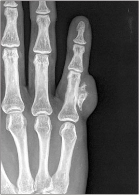
Figure 2 Anteroposterior radiograph taken at this presentation showing a soft tissue mass with radiolucency and an irregularly shaped osseous projection attached to the underlying cortex arising from the lateral aspect of the midshaft of the proximal phalanx of the finger.

Figure 3 Coronal (A) and axial (B) T1-weighted magnetic resonance imaging (MRI) showing a normal, bony signal intensity mass surrounded by a lobulated homogenous soft tissue mass of high signal intensity, coronal (C) and axial (D) T2 weighted MRI showing hypointensity of the soft tissue mass arising from the volar-ulnar aspect of the proximal phalanx shaft.
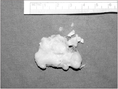
Figure 4 Gross specimen of the excised mass, 3.5×2.5×1.0 cm in size, was well-circumscribed encapsulated fat tissue accompanied by a hard bony mass.

Figure 5 Histopathologically, the superficial area of the mass showed mature lobulated adipose tissue without cellular atypia (A) and the basal area was composed of fibrocartilagenous tissue with immature bony trabeculae (B) (A, B: H&E, ×200).
Reference
-
1. Demos TC, Bruno E, Armin A, Dobozi WR. Parosteal lipoma with enlarging osteochondroma. AJR Am J Roentgenol. 1984; 143:365–366.
Article2. Nora FE, Dahlin DC, Beabout JW. Bizarre parosteal osteochondromatous proliferations of the hands and feet. Am J Surg Pathol. 1983; 7:245–250.
Article3. Yamamoto T, Marui T, Mizuno K. Parosteal lipoma of the distal phalanx: a case report and review of the literature. Clin Orthop Relat Res. 2001; (389):181–184.4. Joseph J, Ritchie D, MacDuff E, Mahendra A. Bizarre parosteal osteochondromatous proliferation: a locally aggressive benign tumor. Clin Orthop Relat Res. 2011; 469:2019–2027.
Article5. Horiguchi H, Sakane M, Matsui M, Wadano Y. Bizarre parosteal osteochondromatous proliferation (Nora's lesion) of the foot. Pathol Int. 2001; 51:816–823.
Article6. Gruber G, Giessauf C, Leithner A, et al. Bizarre parosteal osteochondromatous proliferation (Nora lesion): a report of 3 cases and a review of the literature. Can J Surg. 2008; 51:486–489.7. Chamberlain AM, Anderson KL, Hoch B, Trumble TE, Weisstein JS. Benign parosteal osteochondromatous proliferation of the hand originally diagnosed as osteochondroma: a report of two cases and review. Hand (N Y). 2010; 5:106–110.
Article8. Rajappa S, Kumar MM, Shanmugapriya S. Recurrent solitary osteochondroma of the metacarpal: a case report. J Orthop Surg (Hong Kong). 2013; 21:129–131.
Article9. Rybak LD, Abramovici L, Kenan S, Posner MA, Bonar F, Steiner GC. Cortico-medullary continuity in bizarre parosteal osteochondromatous proliferation mimicking osteochondroma on imaging. Skeletal Radiol. 2007; 36:829–834.
Article10. Ek ET, Slavin JL, Blackney MC, Powell GJ. Parosteal lipoma associated with an underlying osteochondroma arising from the hallux. Skeletal Radiol. 2007; 36:689–692.
Article
- Full Text Links
-
- Actions
-
Cited
- CITED
-
- Close
- Share
-
- Similar articles
-
- A Case of Subungual Bizarre Parosteal Osteochondromatous Proliferation of the Toe
- Bizarre Parosteal Osteochondromatous Proliferation: A Report of One Case
- Bizarre Parosteal Osteochondromatous Proliferation in the First Metatarsal Bone: A Case Report
- Bizarre Parosteal Osteochondromatous Proliferation (Nora's lesion) of the Big Toe. (A Report of Two Cases and Review)
- Bizarre Parosteal Osteochondromatous Proliferation of Middle Phalanx: A Case Report
