J Korean Bone Joint Tumor Soc.
2010 Jun;16(1):37-41. 10.5292/jkbjts.2010.16.1.37.
Juvenile Hemangioma Occurred in Distal Femoral Epiphysis
- Affiliations
-
- 1Department of Orthopedic Surgery, Hanyang University College of Medicine, Seoul, Korea. kimts@hanyang.ac.kr
- 2Department of Orthopedic Surgery, Guri Hospital, Hanyang University College of Medicine, Guri, Korea.
- 3Department of Pathology, Hanyang University College of Medicine, Seoul, Korea.
- KMID: 2006340
- DOI: http://doi.org/10.5292/jkbjts.2010.16.1.37
Abstract
- A hemangioma occurred in the bony epiphysis is extremly rare. A 5-year-old boy visited to our hospital with pain and flexion contracture on the right knee. MRI showed some lesions scattered in the epiphysis of the distal femur and the proximal tibia. Biopsy specimen from the distal femoral epiphysis revealed pathologic findings compatible with hemangioma. On 8 years follow-up, the lesion in the distal femoral epiphysis had been cured, and those in the proximal tibial epiphysis were spontaneously disappeared without surgery. The scanogram shows no leg length discrepancy and angular deformity. We reports a rare case of hemangioma occurred in the bony epiphysis with the results of 8 year follow-up with the review of literatures.
Keyword
MeSH Terms
Figure
-

Figure 1. Anteroposterior (A) and lateral (B) radiographs of the right knee show osteolytic lesion in the distal femoral epiphysis.
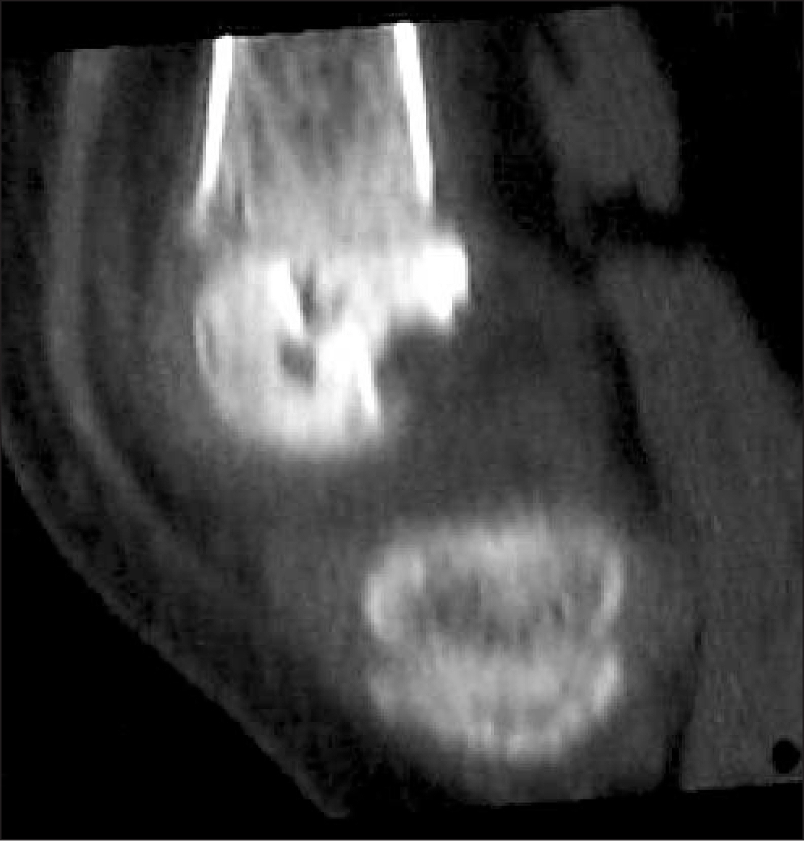
Figure 2. Sagittal CT image shows osteolytic lesion involving distal femoral epiphyseal plate.

Figure 3. Coronal T2-weighted magnetic resonance image shows high signal in tumor.
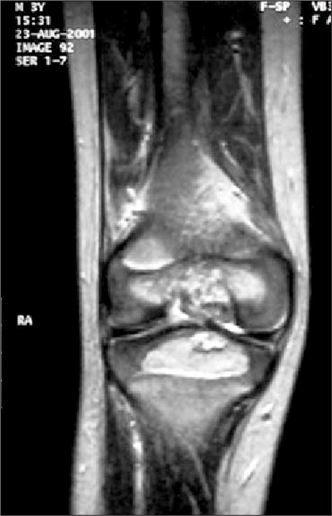
Figure 4. Coronal T1-weighted magnetic resonance image shows heterogenous high signal image in tumor.
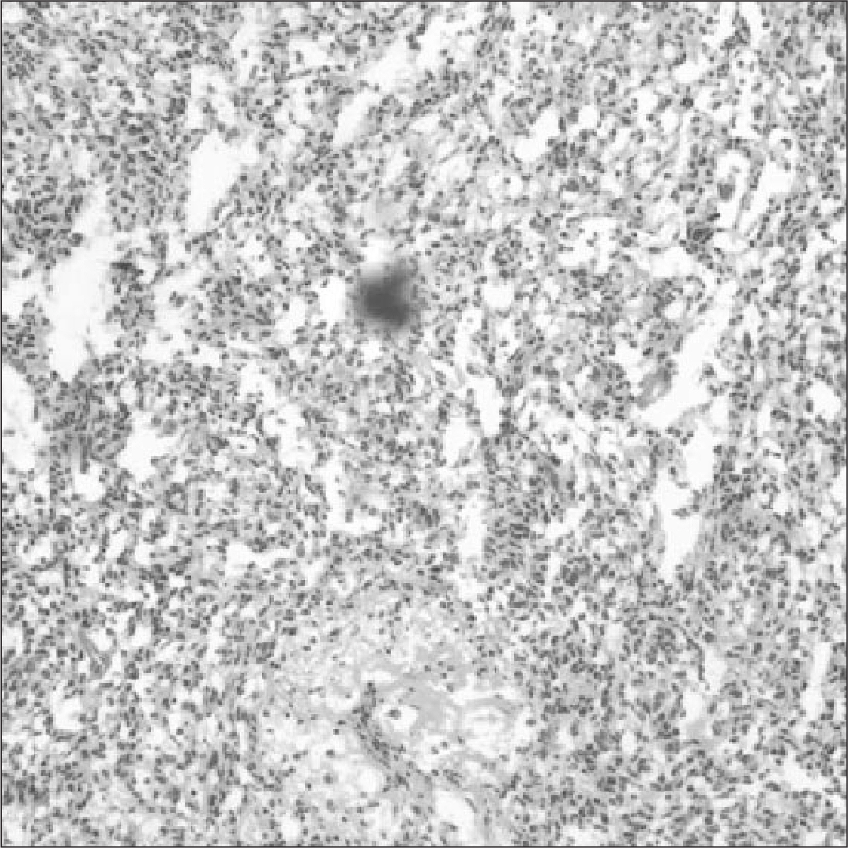
Figure 5. Microscpic finding shows lobular growth of plump endothelial cells lining vascular spaces with small incospicious lumens.
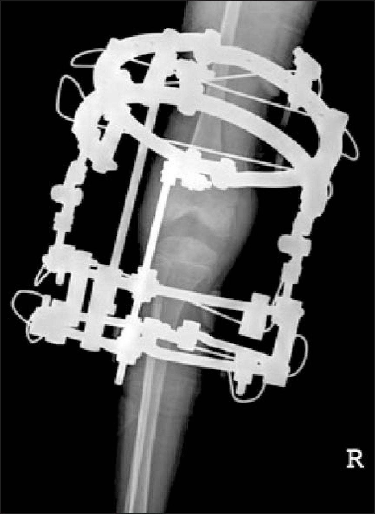
Figure 6. Application of Ilizarov apparatus for flexion contracture.
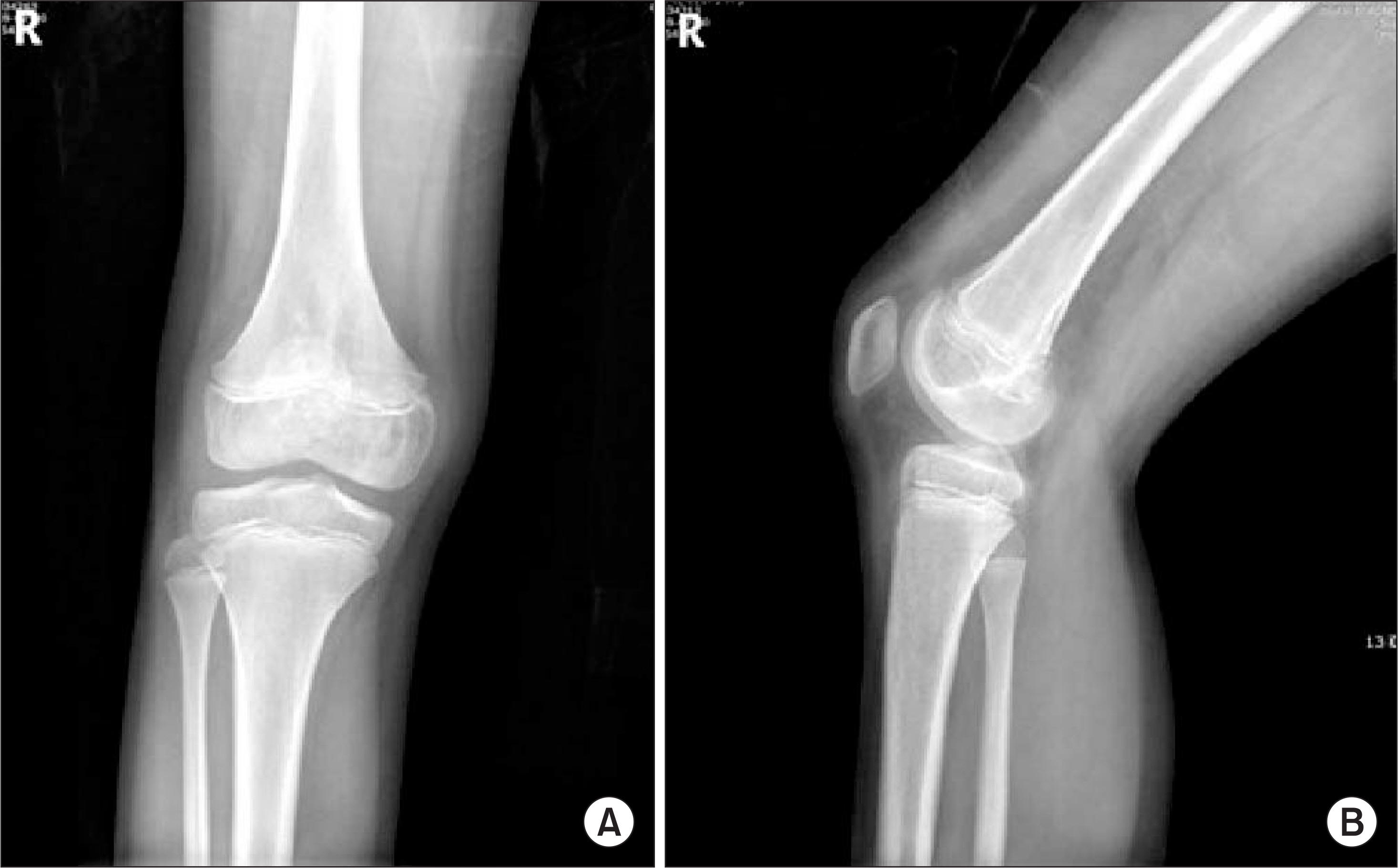
Figure 7. Anteroposterior (A) and lateral (B) radiographs of right knee the 8-year followup show no evidence of hemangioma.
Reference
-
1. Dahlin DC, Unni KK. Introduction and scope of the study in bone tumors: general aspects and data on 8,542 cases. Springfield. 1986; Ill:3–17.2. Kaleem Z, Kyriakos M. Solitary skeletal hemangioma of the extremities. Skeletal Radiol. 2000; 29:502–13.
Article3. Chawla A, Singlakhia M, Maheshwari M, Modi N, Parmar H. Intraosseous hemangioma of the proximal femur: imaging findings. Br J Radio. 2006; 79:64–6.4. Mirra JM. Bone tumors: clinical, radiologic, and pathologic correlations. Philadelphia: Lea & Febiger;1989. p. 1338–81.5. Lopez-Barea F, Harisson D, Lamas M, et al. Intracortical hemangioma of the bone. J Bone and Joint Surg. 1998; 80:1673–8.6. Dorfman HD, Czermiak B. Vascular lesions in bone tumors. St. Louis, MO: Mosby;1998. p. 729–814.7. Stoller DW. Bone and soft tissue tumours. In: MRI in orthopedics and sports medicine. 2nd ed.Lippincot: Williams and Wilkins;1972. p. 1231–339.8. Yochum TR, Rowe LJ. Tumours and tumour like processes. In: Essentials of skeletal radiology. 2nd ed.Williams and Wilkins;1996. p. 975–1192.9. Lee HK, Lee SH, Kim HS, Lee DH, Yang MS. Hemangiomas in soft tissue of trunk and extremities. J Korean Bone Joint Tumor Soc. 1996; 2:178–85.10. Zahid K, Michael K, Wilian GT. Solitary skeletal hemangioma of the extremities. Skeletal Radiol. 2000; 29:502–13.
- Full Text Links
-
- Actions
-
Cited
- CITED
-
- Close
- Share
-
- Similar articles
-
- Pathologic Separation of Capital Femoral Epiphysis due to an Osteosarcoma
- A Clinical Study of the Mechanism of Injury of Juvenile Tillaux Fracture and Triplane Fracture
- Prognostic Factors in Slipped Capital Femoral Epiphysis
- A Clinical Study of Slipped Capital Femoral epiphysis
- Traumatic Separation of Upper Femoral Epiphysis: A Case Report
