J Korean Surg Soc.
2009 Sep;77(3):207-210. 10.4174/jkss.2009.77.3.207.
Extra-gastric Cyst Lesions Mimicking Gastric Submucosal Tumor
- Affiliations
-
- 1Department of Surgery, St. Mary's Hospital, The Catholic University of Korea College of Medicine, Seoul, Korea. kimwook@catholic.ac.kr
- KMID: 2004221
- DOI: http://doi.org/10.4174/jkss.2009.77.3.207
Abstract
- Gastric cancer remains still the most frequent type of cancer despite its declining incidence in Korea. As a result of the health promotion policy of the Korean government and increase in concern for individuals' health, screening endoscopy for detecting early gastric cancer and general physical exams have become widespread. Thereby, the incidental detection of gastric submucosal tumors (SMTs) are now occasionally diagnosed by screening endoscopy. Since endoscopic examination gives little information on SMTs, a variety of studies have been conducted to determine the etiology of SMTs and to distinguish them from extra-luminal compressive lesions. Here, we report one clinical case of cavernous hemangioma on the left lateral section of the liver and one case of omental cyst, which was preoperatively mistaken as gastric SMT.
Keyword
MeSH Terms
Figure
-
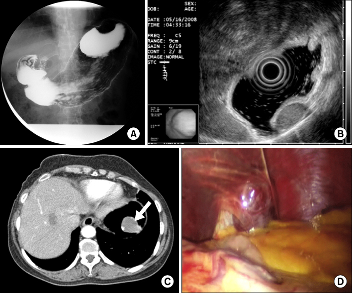
Fig. 1 Barium radiography of stomach shows protruding mass in gastric fundus (A), EUS shows hypoechoic homogenous mass in 4th layer of the gastric wall (B), Abdominal CT shows gastric mass in fundus and arrow indicates hemangioma (C), Cavernous hepatic hemangioma in left lateral section of the liver (D).

Fig. 2 EUS finding suggests hypoechoic cystic mass in 5th layer of the gastric wall (A), Cystic mass located in posterior wall of the stomach (B).
Reference
-
1. Rösch T, Kapfer B, Will U, Baronius W, Strobel M, Lorenz R, et al. Accuracy of endoscopic ultrasonography in upper gastrointestinal submucosal lesions: a prospective multicenter study. Scand J Gastroenterol. 2002. 37:856–862.2. Chak A. EUS in submucosal tumors. Gastrointest Endosc. 2002. 56:S43–S48.3. Chen TK, Wu CH, Lee CL, Lai YC, Yang SS, Tu TC. Endoscopic ultrasonography to study the causes of extragastric compression mimicking gastric submucosal tumor. J Formos Med Assoc. 2001. 100:758–761.4. Ponsaing LG, Kiss K, Loft A, Jensen LI, Hansen MB. Diagnostic procedures for submucosal tumors in the gastrointestinal tract. World J Gastroenterol. 2007. 13:3301–3310.5. Motoo Y, Okai T, Ohta H, Satomura Y, Watanabe H, Yamakawa O, et al. Endoscopic ultrasonography in the diagnosis of extraluminal compressions mimicking gastric submucosal tumors. Endoscopy. 1994. 26:239–242.6. Park JM, Kim J, Kim HI, Kim CS. Hepatic cyst misdiagnosed as a gastric submucosal tumor: a case report. World J Gastroenterol. 2008. 14:3092–3094.7. Lerner SM, Hiatt JR, Salamandra J, Chen PW, Farmer DG, Ghobrial RM, et al. Giant cavernous liver hemangiomas: effect of operative approach on outcome. Arch Surg. 2004. 139:818–821.8. Patriti A, Graziosi L, Sanna A, Gullà N, Donini A. Laparoscopic treatment of liver hemangioma. Surg Laparosc Endosc Percutan Tech. 2005. 15:359–362.9. Conzo G, Vacca R, Grazia Esposito M, Brancaccio U, Celsi S, Livrea A. Laparoscopic treatment of an omental cyst: a case report and review of the literature. Surg Laparosc Endosc Percutan Tech. 2005. 15:33–35.
- Full Text Links
-
- Actions
-
Cited
- CITED
-
- Close
- Share
-
- Similar articles
-
- Gastric Neurilemmoma
- Gastric Pseudotumoral Lesion Caused by a Fish Bone Mimicking a Gastric Submucosal Tumor
- Two cases of mucinous adenocarcinoma of the stomach mistaken as submucosal tumor
- Malignant Solitary Fibrous Tumor of Retroperitoneum Mimicking Gastric Submucosal Tumor
- Comparisons of Gastric Endoscopy and Upper Gastrointestinal Series in The Submucosal Tumor
