Volumetry of Artificial Pulmonary Nodules in Ex Vivo Porcine Lungs: Comparison of Semi-automated Volumetry and Radiologists' Performance
- Affiliations
-
- 1Department of Radiology, Chungnam National University Hospital, Korea. michelan@cnu.ac.kr
- 2Department of Therapeutic Radiology and Oncology, Pusan National University Yangsan Hospital, Korea.
- 3Department of Radiology, Seoul National University Hospital, Korea.
- 4Department of Radiology, Kwandong University College of Medicine, Myungji Hospital, Korea.
- 5Department of Nuclear & Quantum Engineering, KAIST, Korea.
- KMID: 2002952
- DOI: http://doi.org/10.3348/jksr.2010.62.5.447
Abstract
- PURPOSE
With the advent of MSCT, the detection rate of small pulmonary nodules is markedly greater. However, there is no definite diagnostic clue to differentiate between malignant and benign nodules, except for the interval growth in small nodule less than 1 cm in diameter. We evaluated the accuracy of computeraided volumetry (CAV) and compared it with 4 radiologists' measurement.
MATERIALS AND METHODS
Fifteen artificial nodules that were embedded in the ex vivo porcine lung were scanned by MSCT. The diameters and volumes of nodules were independently measured three times, at 5-day intervals, and by four radiologists as well as by CAV. We evaluated the accuracy of the measurements on the basis of the true diameter and volume of the nodules. Using a paired t-test and a Bland-Altman plot, we evaluated whether there was a statistically significant difference between the radiologists' measurements and the CAV.
RESULTS
The accuracy of the manual measurements by radiologists revealed a statistically significant difference from the true diameter and volume of the artificial nodules (p<0.01). Conversely, the accuracy of CAV did not show a statistically significant difference with the true nodule diameter and volume (p>0.01) CONCLUSION: The results of this study suggest that CAV is an accurate and useful tool to evaluate the volume of pulmonary nodules and can eventually be used to differentiate malignant and benign nodules as well as evaluate the therapeutic response of lung cancer.
Figure
-

Fig. 1 Tumor phantoms. Axial image of artificial pulmonary nodules (arrows) in air inflated ex vivo porcine lung.
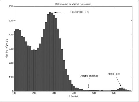
Fig. 2 Histogram for adaptive threshold. Adaptive threshold for each artificial nodule was estimated using mean value of HU value of nodule peak and that of neighborhood peak.

Fig. 3 Graphical user interface of semi-automated volumetry. A. Selected images are shown on the right side of monitor. To start volumetry, click on the selected nodule, then press the volume button. The calculated volume is visible on the ritgt-lower portion of monitor. B. Three-dimensional image from 1 mm thickness MSCT data of an artificial nodule C. The overall workflow for a nodule volume measurement.
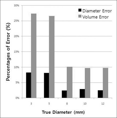
Fig. 4 Relationship between changes in uni-dimensional measurement and tumor volume.

Fig. 5 The percentage of error of the volume estimates by radiologists in 15 synthetic nodules. The percentage of error become greater with decreasing nodule size.
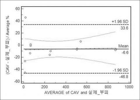
Fig. 6 The percentage of error of the volume estimates by CAV algorithm in 15 synthetic nodules. The percentage of error is nearly fixed at -6.6 % with all nodule.
Reference
-
1. Ko JP, Marcus R, Bomsztyk E, Babb JS, Stefanescu C, Kaur M, et al. Effect of blood vessels on measurement of nodule volume in a chest phantom. Radiology. 2006; 239:79–85.2. WHO. WHO Handbook for Reporting results of cancer treatment. WHO offset publication 48. Geneva, Switzerland: World Health Organization;1979.3. Miller AB, Hoogstraten B, Staquet M, Winkler A. Reportiong results of cancer treatment. Cancer. 1981; 47:207–214.4. Therasse P, Arbuck SG, Eisenhauer EA, Wanders J, Kaplan RS, Rubinstein L, et al. New guidelines to evaluate the response to treatment in solid tumors: european organization for research and treatment of cancer, national cancer institute of the united states, national cancer institute of canada. J Natl Cancer Inst. 2000; 92:205–216.5. Erasmus JJ, Gladish GW, Broemeling L, Sabloff BS, Truong MT, Herbst RS, et al. Interobserver and intraobserver variability in measurement of non-small-cell carcinoma lung lesions: implications for assessment of tumor response. J Clin Oncol. 2003; 21:2574–2582.6. Marten K, Engelke C. Computer-aided detection and automated CT volumetry of pulmonary nodules. Eur Radiol. 2007; 17:888–901.7. Bolte H, Riede C, Müller-Hülsbberg S, Freitag-Wolf S, Kohl G, Drews T, et al. Precision of computer-aided volumetry of artificial small solid pulmonary nodules in ex vivo porcine lungs. Br J Radiol. 2007; 80:414–421.8. Wormanns D, Kohl G, Klotz E, Marheine A, Beyer F, Heindel W, et al. Volumetric measurements of pulmonary nodules at multirow detector CT: in vivo reproducibility. Eur Radiol. 2004; 14:86–92.9. Bolte H, Riedel C, Jahnke T, Inan N, Freitag S, Kohl G, et al. Reproducibility of computer-aided volumetry of artificial small pulmonary nodules in ex vivo porcine lungs. Invest Radiol. 2006; 41:28–35.10. Yankelevitz DF, Reeves AP, Kostis WJ, Zhao B, Henschke CI. Small pulmonary nodules: volumetrically determined growth rates based on CT evaluation. Radiology. 2000; 217:251–256.11. Jennings SG, Winer-Muram HT, Tarver RD, Farber MO. Lung tumor growth: assessment with CT-comparison of diameter and cross-sectional area with volume measurements. Radiology. 2004; 231:866–871.12. Tran LN, Brown MS, Goldin JG, Yan X, Pais RC, McNitt-Gray MF, et al. Comparisons of treatment response classification between unidimensional, bidimensional, and volumetric measurements of metastatic lung lesions on chest computed tomography. Acad Radiol. 2004; 11:1355–1360.13. Schwartz LH, Ginsberg MS, DeCorato D, Rothenberg LN, Einstein S, Kijewski P, et al. Evaluation of tumor measurements in oncology: use of film-based and electronic techniques. J Clin Oncol. 2000; 18:2179–2184.14. Das M, Mühlenbruch G, Katoh M, Bakai A, SalganiCoff M, Stanzel S, et al. Automated volumetry of solid pulmonary nodules in a phantom: accuracy across different CT scanner technologies. Invest Radiol. 2007; 42:297–302.15. Revel MP, Merlin A, Peyrard S, Triki R, Couchon S, Chatellier G, et al. Software volumetric evaluation of doubling times for differentiating benign versus malignant pulmonary nodules. AJR Am J Roentgenol. 2006; 187:135–142.16. Goo JM, Tongdee T, Tongdee R, Yeo K, Hildebolt CF, Bae KT. Volumetric measurement of synthetic lung nodules with multi-detector row CT: effect of various image reconstruction parameters and segmentation thresholds on measurement accuracy. Radiology. 2005; 235:850–856.17. Harris KM, Adams H, Lloyd DC, Harvey DJ. The effect on apparent size of simulated pulmonary nodules of using three standard CT window settings. Clin Radiol. 1993; 47:241–244.18. Van Hoe L, Haven F, Bellon E, Baert AL, Bosmans H, Feron M, et al. Factors influencing the accuracy of volume measurements in spiral CT: a phantom study. J Comput Assist Tomogr. 1997; 21:332–338.19. Honda O, Sumikawa H, Johkoh T, Tomiyama N, Mihara N, Inoue A, et al. Computer-assisted lung nodule volumetry from multi-detector row CT: influence of image reconstruction parameters. Eur J Radiol. 2007; 62:106–113.20. Petrou M, Quint LE, Nan B, Baker LH. Pulmonary nodule volumetric measurement variability as a function of CT slice thickness and nodule morphology. AJR Am J Roentgenol. 2007; 188:306–312.21. Honda O, Johkoh T, Sumikawa H, Inoue A, Tomiyama N, Mihara N, et al. Pulmonary nodules: 3D volumetric measurement with multidetector CT-effect of intravenous contrast medium. Radiology. 2007; 45:881–887.22. Gietema HA, Schaefer-Prokop CM, Mali WP, Groenewegen G, Prokop M. Pulmonary nodules: interscan variability of semiautomated volume measurements with multisection CT--influence of inspiration level, nodule size, and segmentation performance. Radiology. 2007; 245:888–894.
- Full Text Links
-
- Actions
-
Cited
- CITED
-
- Close
- Share
-
- Similar articles
-
- Feasibility of Commercially Available, Fully Automated Hepatic CT Volumetry for Assessing Both Total and Territorial Liver Volumes in Liver Transplantation
- Effect of the High-Pitch Mode in Dual-Source Computed Tomography on the Accuracy of Three-Dimensional Volumetry of Solid Pulmonary Nodules: A Phantom Study
- A Computer-Aided Diagnosis for Evaluating Lung Nodules on Chest CT: the Current Status and Perspective
- Evaluation of Computer Aided Volumetry for Simulated Small Pulmonary Nodules on Computed Tomography
- Clinical implication of hepatic volumetry for living donor liver transplantation
