Pre-operative Evaluation of Consistency in Intra-axial Brain Tumor with Diffusion-weighted Images (DWI) and Conventional MR Images
- Affiliations
-
- 1Department of Radiology, Collage of Medicine, The Catholic University of Korea. ahn-kj@catholic.ac.kr
- KMID: 2000058
- DOI: http://doi.org/10.13104/jksmrm.2011.15.2.102
Abstract
- PURPOSE
To retrospectively evaluate the usefulness of diffusion-weighted images, ADC maps and conventional MR images for determination of brain tumor consistency.
MATERIALS AND METHODS
Twenty-three patients with brain tumor underwent MR examinations with T1, T2 and diffusion-weighted images. Regions of interest (ROIs) were drawn in the tumors, and the measured signal intensities (SI) were normalized with the contralateral side. We evaluated the correlation between SI ratios from various images and tumor consistency assessed at surgery. In three patients with both cystic and solid components, each component was evaluated independently. Qualitatively observed SIs were also correlated with tumor consistency.
RESULTS
Statistical analysis revealed significant correlation between tumor consistency and ADC ratio (r = -0.586, p = 0.002), SI ratios on T2-weighted images (r = -0.497, p = 0.010), and observed SIs on T2-weighted images (r = -0.461, p = 0.018). The relative ratio of ADC value correlated with tumor consistency most strongly.
CONCLUSION
The measured ratio of ADC, SI ratio and observed SI grade on T2-weighted images can provide valuable information about the consistency of brain tumor.
Keyword
Figure
-
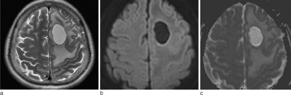
Fig. 1 Metastatic tumor with hard consistency in 44-year-old woman. (a) Axial T2WI shows a homogenous hyperintense (grade 1) mass. (b) Axial DWI shows a hypointense lesion (grade -2) with respect to normal white matter. (c) ADC maps shows a mass with increased diffusion coefficient (grade 1).
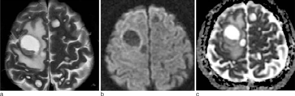
Fig. 2 Metastatic mucoepidermoid carcinoma with gelatinous consistency in 67-year-old man. (a) Axial T2WI shows homogenous hyperintense (grade 3) masses. (b) Axial DWI shows hypointense masses (grade -2) with respect to normal white matter. (c) ADC maps shows masses with increased diffusion coefficient (grade 3).
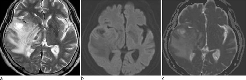
Fig. 3 Glioblastoma with friable consistency in 69-year-old man. (a) Axial T2WI shows a homogenous hyperintense (grade 2) mass. (b) Axial DWI shows a hypointense lesion (grade -1) with respect to normal white matter. (c) ADC maps shows a mass with slightly increased diffusion coefficient (grade 1).
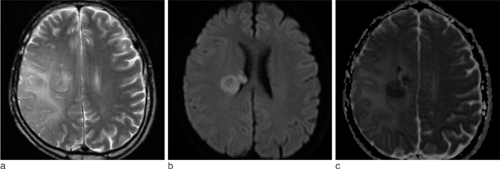
Fig. 4 Glioblastoma with friable consistency in 42-year-old man. (a) Axial T2WI shows a homogenous hyperintense (grade 1) mass. (b) Axial DWI shows a hyperintense lesion (grade 2) with respect to normal white matter. (c) ADC maps shows a mass with slightly decreased diffusion coefficient (grade -2).
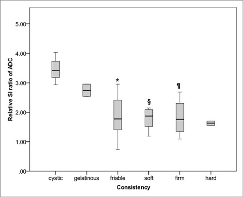
Fig. 5 Box plot shows relative SI ratios on ADC in tumor consistency groups. Relative SI ratios on ADC are significantly correlated with intra-axial brain tumor consistency. *, §, ¶ = significant different from cystic consistency group *p = 0.02 , §p = 0.014, ¶p = 0.032
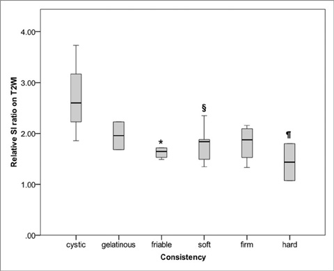
Fig. 6 Box plot shows relative SI ratios on T2WI in tumor consistency groups. Relative SI ratios on T2WI are significantly correlated with intra-axial brain tumor consistency. *, §, ¶ = significant different from cystic consistency group *p = 0.033, §p = 0.044, ¶p = 0.045
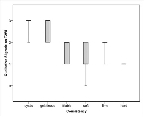
Fig. 7 Box plot shows qualitative SI grade on T2WI in tumor consistency groups. SI grade on T2WI are significantly correlated with intra-axial brain tumor consistency.
Reference
-
1. Iuchi T, Saeki N, Tanaka M, Sunami K, Yamaura A. MRI prediction of fibrous pituitary adenomas. Acta Neurochir (Wien). 1998. 140:779–786.2. Snow RB, Johnson CE, Morgello S, Lavyne MH, Patterson RH Jr. Is magnetic resonance imaging useful in guiding the operative approach to large pituitary tumors? Neurosurgery. 1990. 26:801–803.3. Naganuma H, Satoh E, Nukui H. Technical considerations of transsphenoidal removal of fibrous pituitary adenomas and evaluation of collagen content and subtype in the adenomas. Neurol Med Chir (Tokyo). 2002. 42:202–212. discussion 213.4. Bahuleyan B, Raghuram L, Rajshekhar V, Chacko AG. To assess the ability of MRI to predict consistency of pituitary macroadenomas. Br J Neurosurg. 2006. 20:324–326.5. Chakrabortty S, Oi S, Yamaguchi M, Tamaki N, Matsumoto S. Growth hormone-producing pituitary adenomas: MR characteristics and pre- and postoperative evaluation. Neurol Med Chir (Tokyo). 1993. 33:81–85.6. Hagiwara A, Inoue Y, Wakasa K, Haba T, Tashiro T, Miyamoto T. Comparison of growth hormone-producing and non-growth hormone-producing pituitary adenomas: imaging characteristics and pathologic correlation. Radiology. 2003. 228:533–538.7. Yamaguchi N, Kawase T, Sagoh M, Ohira T, Shiga H, Toya S. Prediction of consistency of meningiomas with preoperative magnetic resonance imaging. Surg Neurol. 1997. 48:579–583.8. Suzuki Y, Sugimoto T, Shibuya M, Sugita K, Patel SJ. Meningiomas: correlation between MRI characteristics and operative findings including consistency. Acta Neurochir (Wien). 1994. 129:39–46.9. Maiuri F, Iaconetta G, de Divitiis O, Cirillo S, Di Salle F, De Caro ML. Intracranial meningiomas: correlations between MR imaging and histology. Eur J Radiol. 1999. 31:69–75.10. Yrjana SK, Tuominen H, Karttunen A, Lahdesluoma N, Heikkinen E, Koivukangas J. Low-field MR imaging of meningiomas including dynamic contrast enhancement study: evaluation of surgical and histopathologic characteristics. AJNR Am J Neuroradiol. 2006. 27:2128–2134.11. Kashimura H, Inoue T, Ogasawara K, et al. Prediction of meningioma consistency using fractional anisotropy value measured by magnetic resonance imaging. J Neurosurg. 2007. 107:784–787.12. Pierallini A, Caramia F, Falcone C, et al. Pituitary macroadenomas: preoperative evaluation of consistency with diffusionweighted MR imaging--initial experience. Radiology. 2006. 239:223–231.13. Suzuki C, Maeda M, Hori K, et al. Apparent diffusion coefficient of pituitary macroadenoma evaluated with line-scan diffusion-weighted imaging. J Neuroradiol. 2007. 34:228–235.14. Mahmoud OM, Tominaga A, Amatya VJ, et al. Role of PROPELLER diffusion-weighted imaging and apparent diffusion coefficient in the evaluation of pituitary adenomas. Eur J Radiol. 2010. (Epub ahead of print).15. Kono K, Inoue Y, Nakayama K, et al. The role of diffusionweighted imaging in patients with brain tumors. AJNR Am J Neuroradiol. 2001. 22:1081–1088.16. Guo AC, Cummings TJ, Dash RC, Provenzale JM. Lymphomas and high-grade astrocytomas: comparison of water diffusibility and histologic characteristics. Radiology. 2002. 224:177–183.17. Gudbjartsson H, Maier SE, Mulkern RV, Morocz IA, Patz S, Jolesz FA. Line scan diffusion imaging. Magn Reson Med. 1996. 36:509–519.18. Pipe JG, Farthing VG, Forbes KP. Multishot diffusion-weighted FSE using PROPELLER MRI. Magn Reson Med. 2002. 47:42–52.
- Full Text Links
-
- Actions
-
Cited
- CITED
-
- Close
- Share
-
- Similar articles
-
- A Comparison of Lesion Detection and Conspicuity on T2-weighted Images (T2 FFE), FLAIR and Diffusion-weighted Images in Patients with Traumatic Brain Injury
- Diffusion Weighted MRI Findings in Two Patients with Hypoxic Brain Damage
- Diffusion-weighted MR Imaging after Intracranial Tumor Resection
- Diffusion-Weighted MR Imaging in Biopsy-Proven Creutzfeldt-Jakob Disease
- MR Imaging Features of Acute Enterovirus 71 Encephalitis in a Patient with Hand-Foot-Mouth Disease: A Case Report
