In-depth morphological study of mesiobuccal root canal systems in maxillary first molars: review
- Affiliations
-
- 1Department of Conservative Dentistry, Kyung Hee University School of Dentistry, Seoul, Korea.
- 2Beautiful Dental Clinic, Changwon, Korea.
- 3Department of Dentistry, Yonsei University Wonju College of Medicine, Wonju, Korea.
- 4Department of Conservative Dentistry, Dental Research Institute and BK 21 Program, Seoul National University Dental Hospital, Seoul National University School of Dentistry, Seoul, Korea. kum6139@snu.ac.kr
- KMID: 1995470
- DOI: http://doi.org/10.5395/rde.2013.38.1.2
Abstract
- A common failure in endodontic treatment of the permanent maxillary first molars is likely to be caused by an inability to locate, clean, and obturate the second mesiobuccal (MB) canals. Because of the importance of knowledge on these additional canals, there have been numerous studies which investigated the maxillary first molar MB root canal morphology using in vivo and laboratory methods. In this article, the protocols, advantages and disadvantages of various methodologies for in-depth study of maxillary first molar MB root canal morphology were discussed. Furthermore, newly identified configuration types for the establishment of new classification system were suggested based on two image reformatting techniques of micro-computed tomography, which can be useful as a further 'Gold Standard' method for in-depth morphological study of complex root canal systems.
Keyword
MeSH Terms
Figure
-

Figure 1 Representative images by (a, c) clearing technique; (b, d) Three-dimensional (3D) volume rendering technique of micro-computed tomography (MCT) showed restricted penetration of dye solution into a root canal system (a, c, white arrows with black outline).
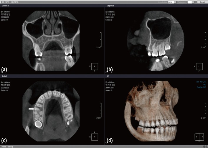
Figure 2 Original cone-beam computed tomography (CBCT) images of a maxillary first molar with two canals in the mesiobuccal (MB) root as viewed coronal, sagittal, axial direction by using OnDemand3D software. (a) Coronal view with the MB root exhibiting Type III canal configuration; (b) Sagittal view; (c) Axial view (arrow indicates the existence of two canals in MB root); (d) Three-dimensional (3D) reconstruction image.

Figure 3 Canal images with different reformatting methods. (a) Simulation of conventional ray projection image; (b) Curved thin slab-minimum intensity projection (TS-MinIP) image; (c) Volume rendered image with segmentation of canal structure; (d) Surface rendered image. TS-MinIP image shows the most clear view of fine canal structures (b, white arrow), while an accessory canal hiding behind main root canal is shown only in three-dimensional (3D) volume rendered image (c, black arrow with white outline).
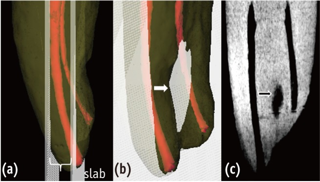
Figure 4 The slab is defined between two planes in (a), and (b) is a rotated image of (a) which shows root surface concavity included in the slab. (c) is a resultant thin slab-minimum intensity projection (TS-MinIP) image with pseudo-canal. If the slab extends beyond the extent of the root due to root concavity or severe curvature (b, white arrow), the pixel value outside the root is recognized as the minimum value and thus can result in a pseudo-canal image (c, black arrow with white outline).
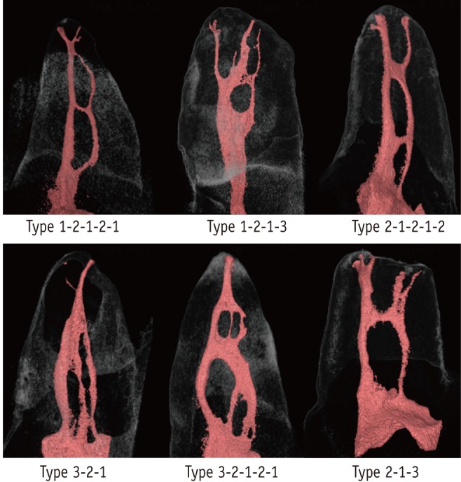
Figure 5 Representative images obtained by three-dimensional rendering which shows six non-classifiable configuration types when the modified Vertucci's classification is applied (Reprinted with the permission of the Clin Oral Investig. Originally published as: Clin Oral Investig 2012, 10.1007/s00784-012-0725-1.).
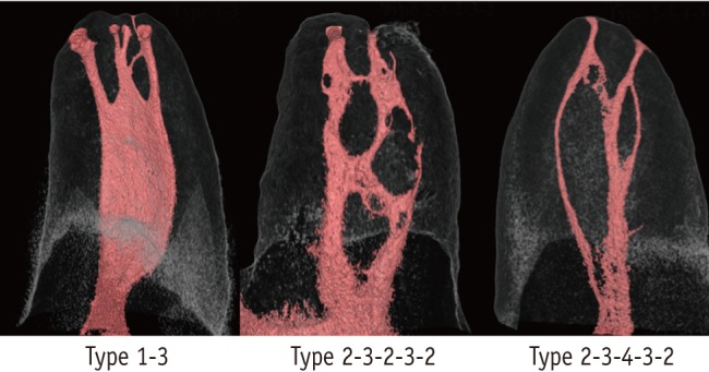
Figure 6 Three-dimensional reconstructed images of 3 non-classifiable configuration types when modified Vertucci's classification was applied. The configuration types were not reported in maxillary first molar MB roots in the literature (Reprinted with the permission of the Clin Oral Investig. Originally published as: Clin Oral Investig 2012, 10.1007/s00784-012-0725-1.).
Reference
-
1. Kim Y, Chang SW, Lee JK, Chen IP, Kaufman B, Jiang J, Cha BY, Zhu Q, Safavi KE, Kum KY. A micro-computed tomography study of canal configuration of multiplecanalled mesiobuccal root of maxillary first molar. Clin Oral Investig. 2012; 10. [Epub ahead of print]. doi:10.1007/s00784-012-0852-8.
Article2. Versiani MA, Pécora JD, de Sousa-Neto MD. Root and root canal morphology of four-rooted maxillary second molars: a micro-computed tomography study. J Endod. 2012; 38:977–982. PMID: 22703664.
Article3. Du Y, Soo I, Zhang CF. A case report of six canals in a maxillary first molar. Chin J Dent Res. 2011; 14:151–153. PMID: 22319758.4. Badole GP, Bahadure RN, Warhadpande MM, Kubde R. A rare root canal configuration of maxillary second molar: a case report. Case Rep Dent. 2012; 2012:767582. doi: 10.1155/2012/767582. Epub 2012 Jul 8. PMID: 22830061.
Article5. Spagnuolo G, Ametrano G, D'Antò V, Formisano A, Simeone M, Riccitiello F, Amato M, Rengo S. Microcomputed tomography analysis of mesiobuccal orifices and major apical foramen in first maxillary molars. Open Dent J. 2012; 6:118–125. PMID: 22905069.
Article6. Kim Y, Lee SJ, Woo J. Morphology of maxillary first and second molars analyzed by cone-beam computed tomography in a Korean population: variations in the number of roots and canals and the incidence of fusion. J Endod. 2012; 38:1063–1068. PMID: 22794206.
Article7. Abuabara A, Baratto-Filho F, Aguiar Anele J, Leonardi DP, Sousa-Neto MD. Efficacy of clinical and radiological methods to identify second mesiobuccal canals in maxillary first molars. Acta Odontol Scand. 2013; 71:205–209. PMID: 22320229.
Article8. Peeters HH, Suardita K, Setijanto D. Prevalence of a second canal in the mesiobuccal root of permanent maxillary first molars from an Indonesian population. J Oral Sci. 2011; 53:489–494. PMID: 22167035.
Article9. Villas-Bôas MH, Bernardineli N, Cavenago BC, Marciano M, Del Carpio-Perochena A, de Moraes IG, Duarte MH, Bramante CM, Ordinola-Zapata R. Micro-computed tomography study of the internal anatomy of mesial root canals of mandibular molars. J Endod. 2011; 37:1682–1686. PMID: 22099905.
Article10. Verma P, Love RM. A Micro CT study of the mesiobuccal root canal morphology of the maxillary first molar tooth. Int Endod J. 2011; 44:210–217. PMID: 20880136.
Article11. Jung IY, Seo MA, Fouad AF, Spångberg LS, Lee SJ, Kim HJ, Kum KY. Apical anatomy in mesial and mesiobuccal roots of permanent first molars. J Endod. 2005; 31:364–368. PMID: 15851930.
Article12. Boruah LC, Bhuyan AC. Morphologic characteristics of root canal of mandibular incisors in North-East Indian population: an in vitro study. J Conserv Dent. 2011; 14:346–350. PMID: 22144800.13. Jain A, Bahuguna R. Root canal morphology of mandibular first premolar in a gujarati population - an in vitro study. Dent Res J (Isfahan). 2011; 8:118–122. PMID: 22013473.14. Weine FS, Healey HJ, Gerstein H, Evanson L. Canal configuration in the mesiobuccal root of the maxillary first molar and its endodontic significance. Oral Surg Oral Med Oral Pathol. 1969; 28:419–425. PMID: 5257186.
Article15. De Vos W, Casselman J, Swennen GR. Cone-beam computerized tomography (CBCT) imaging of the oral and maxillofacial region: a systematic review of the literature. Int J Oral Maxillofac Surg. 2009; 38:609–625. PMID: 19464146.
Article16. Neelakantan P, Subbarao C, Abuja R, Subbarao CV, Gutmann JL. Cone-beam computed tomography study of root and canal morphology of maxillary first and second molars in an Indian population. J Endod. 2010; 36:1622–1627. PMID: 20850665.
Article17. Ritman EL. Current status of developments and applications of micro-CT. Annu Rev Biomed Eng. 2011; 13:531–552. PMID: 21756145.
Article18. Gu Y, Lee JK, Spångberg LS, Lee Y, Park CM, Seo DG, Chang SW, Hur MS, Hong ST, Kum KY. Minimum-intensity projection for in-depth morphologic study of mesiobuccal root. Oral Surg Oral Med Oral Pathol Oral Radiol Endod. 2011; 112:671–677. PMID: 21821440.19. Neelakantan P, Subbarao C, Ahuja R, Subbarao CV. Root and canal morphology of Indian maxillary premolars by a modified root canal staining technique. Odontology. 2011; 99:18–21. PMID: 21271321.
Article20. Yoshioka T, Villegas JC, Kobayashi C, Suda H. Radiographic evaluation of root canal multiplicity in mandibular first premolars. J Endod. 2004; 30:73–74. PMID: 14977299.
Article21. Weng XL, Yu SB, Zhao SL, Wang HG, Mu T, Tang RY, Zhou XD. Root canal morphology of permanent maxillary teeth in the Han nationality in Chinese Guanzhong area: a new modified root canal staining technique. J Endod. 2009; 35:651–656. PMID: 19410077.
Article22. Seo MS, Park DS. C-shaped root canals of mandibular second molars in a Korean population: clinical observation and in vitro analysis. Int Endod J. 2004; 37:139–144. PMID: 14871181.23. Cotton TP, Geisler TM, Holden DT, Schwartz SA, Schindler WG. Endodontic applications of cone-beam volumetric tomography. J Endod. 2007; 33:1121–1132. PMID: 17931947.
Article24. Michetti J, Maret D, Mallet JP, Diemer F. Validation of cone beam computed tomography as a tool to explore root canal anatomy. J Endod. 2010; 36:1187–1190. PMID: 20630296.
Article25. Lee JH, Kim KD, Lee JK, Park W, Jeong JS, Lee Y, Gu Y, Chang SW, Son WJ, Lee WC, Baek SH, Bae KS, Kum KY. Mesiobuccal root canal anatomy of Korean maxillary first and second molars by cone-beam computed tomography. Oral Surg Oral Med Oral Pathol Oral Radiol Endod. 2011; 111:785–791. PMID: 21439860.
Article26. Zheng Q, Zhang L, Zhou X, Wang Q, Wang Y, Tang L, Song F, Huang D. C-shaped root canal system in mandibular second molars in a Chinese population evaluated by cone-beam computed tomography. Int Endod J. 2011; 44:857–862. PMID: 21599707.
Article27. Lofthag-Hansen S, Huumonen S, Gröndahl K, Gröndahl HG. Limited cone-beam CT and intraoral radiography for the diagnosis of periapical pathology. Oral Surg Oral Med Oral Pathol Oral Radiol Endod. 2007; 103:114–119. PMID: 17178504.
Article28. Blattner TC, George N, Lee CC, Kumar V, Yelton CD. Efficacy of cone-beam computed tomography as a modality to accurately identify the presence of second mesiobuccal canals in maxillary first and second molars: a pilot study. J Endod. 2010; 36:867–870. PMID: 20416435.
Article29. La SH, Jung DH, Kim EC, Min KS. Identification of independent middle mesial canal in mandibular first molar using cone-beam computed tomography imaging. J Endod. 2010; 36:542–545. PMID: 20171380.
Article30. de Paula-Silva FW, Wu MK, Leonardo MR, da Silva LA, Wesselink PR. Accuracy of periapical radiography and cone-beam computed tomography scans in diagnosing apical periodontitis using histopathological findings as a gold standard. J Endod. 2009; 35:1009–1012. PMID: 19567324.31. Wu M-K, Wesselink PR, Shemesh H, Patel S. Endodontic epidemiologic investigations and clinical outcome studies with cone-beam computed tomography. J Endod. doi: 10.1016/j.joen.2011.03.019. [published online 2011 May 19].
Article32. White SC, Pharoah MJ. Oral radiology: principles and interpretation. 2009. 6th ed. St. Louis: Mosby/Elsevier.33. Patel S, Dawood A, Ford TP, Whaites E. The potential applications of cone beam computed tomography in the management of endodontic problems. Int Endod J. 2007; 40:818–830. PMID: 17697108.
Article34. Kim HC, Park SJ, Park SI, Park SH, Kim HJ, Shin HC, Bae WK, Kim IY, Lee HK. Multislice CT cholangiography using thin-slab minimum intensity projection and multiplanar reformation in the evaluation of patients with suspected biliary obstruction: preliminary experience. Clin Imaging. 2005; 29:46–54. PMID: 15864842.35. Satoh S, Ohdama S, Shibuya H. Sliding thin slab, minimum intensity projection imaging for objective analysis of emphysema. Radiat Med. 2006; 24:415–421. PMID: 16958422.
Article36. Bhalla M, Naidich DP, McGuinness G, Gruden JF, Leitman BS, McCauley DI. Diffuse lung disease: assessment with helical CT-preliminary observations of the role of maximum and minimum intensity projection images. Radiology. 1996; 200:341–347. PMID: 8685323.
Article37. Endo S, Murayama F, Hasegawa T, Sohara Y, Fuse K. Helical computed tomographic minimum-intensity projection of a slit in an airway obstruction. Ann Thorac Surg. 1999; 67:847–849. PMID: 10215248.
Article38. Lorensen WE, Cline HE. Marching cubes: a high resolution 3D surface construction algorithm. Comput Graph. 1987; 21:163–169.
Article39. Elvins TT. A survey of algorithms for volume visualization. Comput Graph. 1992; 26:194–201.
Article40. Levoy M. Display of surfaces from volume data. IEEE Comput Graph Appl. 1988; 8:29–37.
Article41. Schroeder WJ, Zarge JA, Lorensen WE. Decimation of triangle meshes. Comput Graph. 1992; 26:65–70.
Article42. Vertucci FJ. Root canal anatomy of the human permanent teeth. Oral Surg Oral Med Oral Pathol. 1984; 58:589–599. PMID: 6595621.
Article43. Ng YL, Aung TH, Alavi A, Gulabivala K. Root and canal morphology of Burmese maxillary molars. Int Endod J. 2001; 34:620–630. PMID: 11762499.
Article44. Park JW, Lee JK, Ha BH, Choi JH, Perinpanayagam H. Three-dimensional analysis of maxillary first molar mesiobuccal root canal configuration and curvature using micro-computed tomography. Oral Surg Oral Med Oral Pathol Oral Radiol Endod. 2009; 108:437–442. PMID: 19386518.
Article45. Somma F, Leoni D, Plotino G, Grande NM, Plasschaert A. Root canal morphology of the mesiobuccal root of maxillary first molars: a micro-computed tomographic analysis. Int Endod J. 2009; 42:165–174. PMID: 19134045.
Article46. Zheng QH, Wang Y, Zhou XD, Wang Q, Zheng GN, Huang DM. A cone-beam computed tomography study of maxillary first permanent molar root and canal morphology in a Chinese population. J Endod. 2010; 36:1480–1484. PMID: 20728713.
Article47. Thomas RP, Moule AJ, Bryant R. Root canal morphology of maxillary permanent first molar teeth at various ages. Int Endod J. 1993; 26:257–267. PMID: 8300257.
Article48. Eder A, Kantor M, Nell A, Moser T, Gahleitner A, Schedle A, Sperr W. Root canal system in the mesiobuccal root of the maxillary first molar: an in vitro comparison study of computed tomography and histology. Dentomaxillofac Radiol. 2006; 35:175–177. PMID: 16618851.
- Full Text Links
-
- Actions
-
Cited
- CITED
-
- Close
- Share
-
- Similar articles
-
- Morphological characteristics of the mesiobuccal root in the presence of a second mesiobuccal canal: a micro-CT study
- Assessment of Root and Root Canal Morphology of Human Primary Molars using CBCT
- Dilemmas pertaining to three canals in the mesiobuccal root of a maxillary second molar: a case report
- Apical periodontitis in mesiobuccal roots of maxillary molars: influence of anatomy and quality of root canal treatment, a CBCT study
- Assessment of the relationship between the maxillary molars and adjacent structures using cone beam computed tomography
