Immunohistochemical study on the distribution of ion channels in rat trigeminal sensory nucleus
- Affiliations
-
- 1Department of Conservative Dentistry, College of Dentistry, Kyung Hee University, Korea.
- KMID: 1987280
- DOI: http://doi.org/10.5395/JKACD.2002.27.3.215
Abstract
- No abstract available.
MeSH Terms
Figure
-

Fig. 1(a) A cell stained with sodium channel antibodies in the trigeminal spinal nucleus (indicated with an arrow)
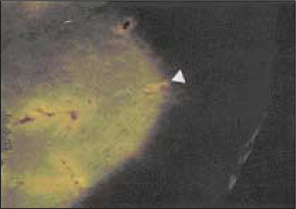
Fig. 1(b) A fluorescence micrograph showing stained sodium channels with sodium channel antibodies in the trigeminal spinal nucleus (indicated with an arrow)

Fig. 2(a) A cell stained with sodium channel antibodies in the trigeminal principal nucleus (indicated with an arrow)
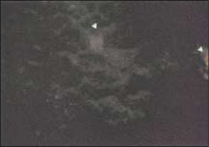
Fig. 2(b) A fluorescence micrograph showing stained sodium channels with sodium channel antibodies in the trigeminal principal nucleus (indicated with an arrow)
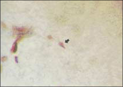
Fig. 3(a) A cell stained with N-type calcium channel antibodies in the trigeminal spinal nucleus (indicated with an arrow)
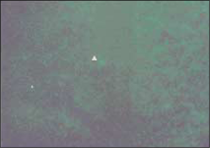
Fig. 3(b) A fluorescence micrograph showing stained N-type calcium channels with calcium channel antibodies in the trigeminal spinal nucleus (indicated with an arrow)

Fig. 4(a) A cell stained with N-type calcium channel antibodies in the trigeminal principal nucleus (indicated with an arrow)

Fig. 4(b) A fluorescence micrograph showing stained N-type calcium channels with calcium channel antibodies in the trigeminal principal nucleus (indicated with an arrow)

Fig. 5(a) A cell stained with P/Q-type calcium channel antibodies in the trigeminal spinal nucleus (indicated with an arrow)
Fig. 5(b) A fluorescence micrograph showing stained P/Q-type calcium channels with calcium channel antibodies in the trigeminal spinal nucleus (indicated with an arrow)

Fig. 6(a) A cell stained with P/Q-type calcium channel antibodies in the trigeminal principal nucleus (indicated with an arrow)
Fig. 6(b) A fluorescence micrograph showing stained P/Q-type calcium channels with calcium channel antibodies in the trigeminal principal nucleus (indicated with an arrow)

Fig. 7(a) A cell stained with R-type calcium channel antibodies in the trigeminal spinal nucleus (indicated with an arrow)
Fig. 7(b) A fluorescence micrograph showing stained R-type calcium channels with calcium channel antibodies in the trigeminal spinal nucleus (indicated with an arrow)

Fig. 8(a) A cell stained with R-type calcium channel antibodies in the trigeminal principal nucleus (indicated with an arrow)
Fig. 8(b) A fluorescence micrograph showing stained R-type calcium channels with calcium channel antibodies in the trigeminal principal nucleus (indicated with an arrow)
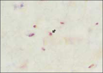
Fig. 9(a) A cell stained with Kir 2.1 potassium channel antibodies in the trigeminal spinal nucleus (indicated with an arrow)

Fig. 9(b) A fluorescence micrograph showing stained potassium channels with Kir 2.1 potassium channel antibodies in the trigeminal spinal nucleus (indicated with an arrow)

Fig. 10(a) A cell stained with Kir 2.1 potassium channel antibodies in the trigeminal principal nucleus (indicated with an arrow)

Fig. 10(b) A fluorescence micrograph showing stained potassium channels with Kir 2.1 potassium channelantibodies in the trigeminal principal nucleus (indicated with an arrow)
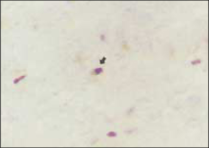
Fig. 11(a) A cell stained with Kv 4.2 potassium channel antibodies in the trigeminal spinal nucleus (indicated with an arrow)
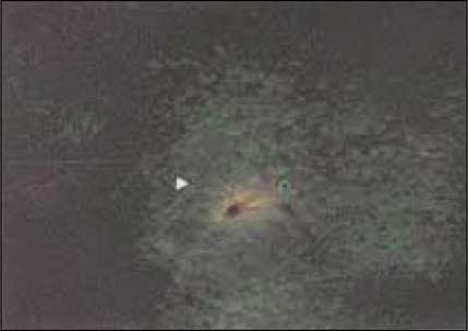
Fig. 11(b) A fluorescence micrograph showing stained potassium channels with Kv 4.2 potassium channel antibodies in the trigeminal spinal nucleus (indicated with an arrow)

Fig. 12(a) A cell stained with Kv 4.2 potassium channel antibodies in the trigeminal principal nucleus (indicated with an arrow)
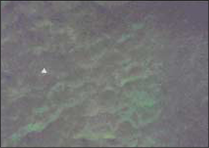
Fig. 12(b) A fluorescence micrograph showing stained potassium channels with Kv 4.2 potassium channel antibodies in the trigeminal principal nucleus (indicated with an arrow)
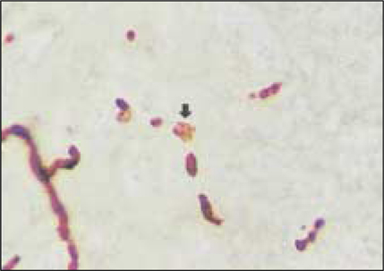
Fig. 13(a) A cell stained with BKCa potassium channel antibodies in the trigeminal spinal nucleus (indicatedwith an arrow)
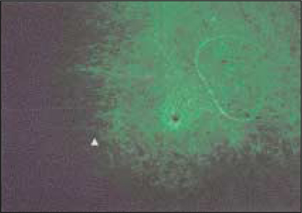
Fig. 13(b) A fluorescence micrograph showing stained potassium channels with BKCa potassium channel antibodies in the trigeminal spinal nucleus (indicated with an arrow)
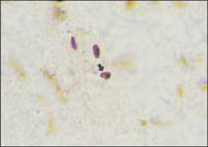
Fig. 14(a) A cell stained with BKCa potassium channel antibodies in the trigeminal principal nucleus (indicated with an arrow)

Fig. 14(b) A fluorescence micrograph showing stained potassium channels with BKCa potassium channel antibodies in the trigeminal principal nucleus (indicated with an arrow)
Reference
-
1. AGNEW WS, MOORE AC, LEVINSON SR, RAFTERY MA. Identification of a large molecular weight peptide associated with a tetrodotoxin binding proteins from the electroplax of Electrophorus electricus. Biochem Biophys Res Commun. 1980. 92:860–866.
Article2. Ahlijanian MK, Westenbroek RE, Catterall WA. Subunit structure and localization of dihydropyridine-sensitive calcium channels in mammalian btain, spinal cord, retina. Neuron. 1990. 4:819–832.
Article3. Alonso G, Widmer H. Clustering of Kv4.2 potassium channels in Phostsy. 1997.
Article4. ALVARADO-MALLART RM, BATINI C, BUISSERET-DELMAS C, CORVISER J. Trigeminal representations of the masticatory and extraocular proprioceptors as revealed by horseradish perioxidase retrograde transport. Exp Brain Res. 1975. 23:167–179.
Article5. APPEL SH, ENGELHARDT JI, BARCIA J, STEFANI E. Immunoglobulins from animal models of motor neuron disease and from human amyotrophic lateral sclerosis patients passively transfer physiological abnormalities to the neuromuscular junction. Proc Natl Acad Sci USA. 1991. 88:647–651.
Article6. APPEL SH, STEFANI E. Amyotrophic lateral sclerosis : etiology and pathogenesis. Curr Neurol. 1991. 11:287–310.7. ARMSTRONG CM. Sodium channels and gating currents. Physiol Rev. 1981. 61:644–682.
Article8. BARCHI RL. Protein components of the purified sodium channel from rat skeletal sarcolemma. J Neurochem. 1983. 36:1377–1385.9. Barry DM, Xu H, Schuessler RB, Nerbonne JM. Functional knockout of the transient outward current, long-QT syndrome, cardiac remodeling in mice expressing a dominant-negative Kv4 alpha subunit. Circ Res. 1998. 83:560–567.
Article10. Bean BP. Classes of calcium channels in vertebrate cells. Annu Rev Physiol. 1989. 51:367–384.
Article11. BENESKI DA, CATTERALL WA. Covalent labeling of protein components of the sodium channel with a photoactivable derivative of scorpion toxin. Proc Natl Acad Sci U S A. 1980. 77:639–643.
Article12. BIERVERT C, SCHROEDER BC, KUBISCH C, BERKOVIC SF, PROPPING P, JENTSCH TJ, STEINLEIN OK. A potassium channel mutation in neonatal human epilepsy. Science. 1998. 279:403–406.
Article13. BROWNE KL, GANCHER ST, NUTT JG, BRUNT ER, SMITH EA, KRAMER P, LITT M. Episodic ataxia/myokymia syndrome is associated with point mutations in the human potassium channel gene KCNA1. Nat Genet. 1994. 8:136–140.
Article14. BUISSERET-DELMAS C, BUISSERET P. Central projections of extraocular muscle afferents in cat. Neurosci Lett. 1990. 109:48–53.
Article15. CATTERALL WA. Neurotoxins that act on voltagesensitive sodium channels in excitable membranes. Annu Rev Pharmacol Toxicol. 1980. 20:15–43.
Article16. CODY FWJ, LEE RWH, TAYLOR AA. functional analysis of the components of the mesencephalic nucleus of the fifth nerve in the cat. J Physiol (London). 1972. 226:249–261.
Article17. Cooper K, Rae JL, Deway J. Inwardly rectifying potassium current in mammalian lens epithelial cells. Am J Physiol. 1991. 261:C115–C123.
Article18. COPRAY JCVM, TERHORST GJ, LIEM RSB, VAN WILLINGEN JD. Neurotransmitters and neuropeptides within the mesencepalic trigeminal nucleus of the rat : an immunohistochemical analysis. Neuroscience. 1990. 37:399–411.
Article19. DEZEEUW CI, BERREBI AS. Postsynaptic targets of Purkinje cell terminals in the cerebellar and vestibular nuclei of the rat. Eur J Neurosci. 1995. 7:2322–2333.
Article20. DELBONO O, GARCIA J, APPEL SH, STEFANI E. Ca2+ current and charge movement of mammalian muscle : action of amyotrophic lateral sclerosis immunoglobulins. J Physiol. 1991. 444:723–742.
Article21. DELBONO O, MAGNELLI V, SAWADA T, SMITH RG, APPEL SH, STEFANI E. Fab fragments from amyotrophic lateral sclerosis IgG affect Ca2+ channels of skeletal muscle. Am J Physiol. 1993. 264:C537–C543.22. DESSEM D, TAYLOR A. Morphology of jawmuscle spindle afferents in the rat. J Comp Neurol. 1989. 282:389–403.
Article23. Doughty JM, Barnes-Davies M, Rusznak Z, Harasztosi C, Forsythe ID. Constructing Ca2+ channel subtypes at cell bodies and synaptic terminals of rat anterioventral cochlear bushy neurons. J Physiol. 1998. 512:365–376.
Article24. Dunlap K, Luebke JI, Turner TJ. Exocytotic Ca2+ channels in mammalian central neurons. Trends Neurosci. 1995. 18:89–98.
Article25. ENGEL AG. Review of evidence for loss of motor nerve terminal Ca2+ channels in Lambert-Eaton myasthenic syndrome. Ann NY Acad Sci. 1991. 635:246–258.
Article26. Fox AP, Nowycky MC, Tsien RW. Kinetic and pharmacological properties distinguishing three types of calcium currents in chick sensory neurones. J Physiol Lond. 1987(a). 394:149–172.
Article27. Fox AP, Nowycky MC, Tsien RW. Single-channel recordings of three types of calcium channels in chick sensory neurones. J Physiol Lond. 1987(b). 394:173–200.
Article28. Galdzicki Z, Puia G, Sciancalepore M, Moran O. Voltage-dependent calcium currents in trigeminal chick neurons. Biochem Biophys Res Comm. 1990. 167:1015–1021.
Article29. GOBEL S, FALLS WM, HOCKFIELD S. Anderson DJ, Matthews B, editors. The division of the dorsal and ventral horns of the mammalian caudal medulla eight layers using anatomical criteria. Pain in the trigeminal region. 1977. Amsterdam: Elsevier;443–453.30. HARTSHORNE RP, CATTERALL WA. Purification of the saxitoxin receptor of the sodium channel from rat brain. Proc Natl Acad Sci USA. 1981. 78:4620–4624.
Article31. HARTSHORNE RP, MESSNER DJ, COPERSMITH JC, CATTERALL WA. The saxitation receptor of the sodium channel from rat brain. Evidence for two nonidentical beta subunits. J Biol Chem. 1982. 257:13888–13891.
Article32. Haydon PG, Henderson E, Stanley E. Localization of individual calcium channels at the release face of a presynaptic nerve terminal. Neuron. 1994. 13:1275–1280.
Article33. HILLE B. Ionic channels of excitable membranes. 1984. first edition. Sunderland, MA: Sinauer Associates, Inc.34. Hille B. Ionic channels of Excitable Membranes. 1992. Sunderland, MA: Sinauer.35. Hodgkin AL, Huxley AF. Currents carried by sodium and potassium ions through the membrane of the giant axon of Loligo. J Physiol Lond. 1952. 116:449–472.
Article36. Hoffman DA, Magee JC, Colbert CM, Johnston D. K+ channel regulation of signal propagation in dendrites of hippocampal pyramidal neurons. Nature. 1997. 387:869–875.
Article37. Jan LY, Jan YN. Cloned potassium channels from eukaryotes and prokaryotes. Annu Rev Neurosci. 1997. 20:91–123.
Article38. JERGE CR. Organization and function of the trigeminal mesencephalic nucleus. J Neurophysiol. 1963. 26:379–392.
Article39. Josephson IR. Properties of inwardly rectifying K+ channels in venricular myocytes. Mol Cell Biochem. 1988. 80:21–26.
Article40. Kaczorowski GJ, Knaus HG, Leonard RJ, Mcmanus OB, Garcia ML. High-conductance calcium-activated potassium channels : structure, pharmacology, function. J Bioenerg Biomembr. 1996. 28:255–267.
Article41. KIM YI. Passive transfer of the Lambert-Eaton myasthenic syndrome : neuromuscular transmission. Muscle Nerve. 1985. 8:162–172.
Article42. LANG B, NEWSOM-DAVIS J, PRIOR C, WRAY PW. Antibodies to motor-nerve terminals : an electrophysiological study of a human myasthenic syndrome transferred to mouse. J Physiol. 1983. 344:335–345.
Article43. LIEM RSB, COPRAY JCVM, VANWILLIGEN JD. Distribution of synaptic boutons in the mesencephalic trigeminal nucleus of the rat. A quantitative electron-microscopical study. Acta Anat (Basel). 1992. 143:74–78.
Article44. Llinas R, Sugimory M, Lin JW, Cherksey B. Blocking and isolation of a calcium channel from neurons in mammals and cephalopods utilizing a toxin fraction FTX from funnel-web spider poison. Proc Natl Acad Sci USA. 1989. 86:5656–5660.45. Maletic-Savatic M, Lenn NJ, TRIMMER JS. Differential spatiotemporal expression of K+ channel polypeptides in rat hippocampal neurons developing in situ and in vitro. J Neurosci. 1995. 15:3840–3851.
Article46. Marty A. The physiological role of calcium-dependent channels. Trends Neurosci. 1989. 12:420–424.
Article47. MATESZ C. Peripheral and central distribution of fibres of the mesencephalic trigeminal root in the rat. Neurosci Lett. 1981. 27:13–17.
Article48. MILLER JA, AGNEW WS, LEVINSON SR. Principal glycopeptide of the tetrodotoxin/saxitoxin binding protein from Electrophorous electricus : isolation and partial chemical and physical characterization. Biochemistry. 1983. 22:462–470.
Article49. Miller RJ. Receptor-mediated regulation of calcium channels and neurotransmitter release. FASEB J. 1990. 4:3291–3299.
Article50. Mintz I, et al. P type calcium channels in rat central and peripheral neurons. Neuron. 1992. 9:85–95.
Article51. MORTON ME, CASSIDY TN, FROEHNER SC, GILMOUR BP, LAURENS RL. Alpha 1 and alpha 2 Ca2+ channel subunit expression in human neuronal and small cell carcinoma cells. FASEB J. 1994. 8:884–888.
Article52. Nelson MT, Cheng H, Rubart M, Santana LF, Bonev AD, Knot HJ, Lederer WJ. Relaxation of arterial smooth muscle by calcium sparks. Science. 1995. 270:633–637.
Article53. Nilius B, Schwarz G, Droogmans G. Modulation by histamine of an inwardly rectifying potassium channel in huma endotherial cells. J Physiol. 1993. 472:359–371.
Article54. Nowycky MC, Fox AP, Trien RW. Three types of neuronal calcium channel with different calcium agonist sensitivity. Nature Lond. 1985. 316:440–443.
Article55. OLSZEWSKI J. On the anatomical and functional organization of the trigeminal nucleus. J Comp Neurol. 1950. 92:401–413.
Article56. PHELAN KD, FALLS WM. An analysis of the cyto- and myeloarchitectonic organization of trigeminal nucleus interpolars in the rat. Somatosens Motor Res. 1989. 6:333–366.
Article57. Puil E, Gimbarzevsky B, Miura RM. Quantification of membrane properties of trigeminal root gangion neurons in guinea pigs. J Neurophysiol. 1986. 55:995–1016.
Article58. Randall A, Tsien R. Pharmacological dissection of multiple types of calcium channel currents in rat cerebellar granule neurons. J Neurosci. 1992. 15(4):2995–3012.
Article59. Regan LJ, Sah DWY, Bean BP. aCalcium channels in rat central and peripheral neurons : high-threshold current resistant to dihydropyridine blockers and w-conotoxin. Neuron. 1991. 6:269–280.
Article60. Regehr WG, Mintz IM. Participation of multiple calcium channel types in transmission at single climbing fiber to Purkinje cell synapses. Neuron. 1994. 12:605–613.
Article61. RENEHAN WE, JACQUIN MF. Olesen J, Tfelt-Hansen P, Welch K, editors. Anatomy of central nervous system pathways related to head pain. The headaches. 1993. New York: Raven Press;59–68.62. RITCHIE JM, ROGART RB. The binding of saxitoxin and thetrodotoxin to excitable tissue. Rev Physiol Biochem Pharmacol. 1977. 79:1–49.63. ROWL LP. Amyotrophic lateral sclerosis : theories and therapies. Ann Neurol. 1994. 35:129–130.64. Scott RH, Pearson HA, Dolphin AC. Aspects of vertebrate neuronal voltage-activated calcium currents and their regulation. Prog Neurobiol. 1991. 36:485–520.
Article65. Scroggs RS, Fox AP. Calcium current variation between acutely isolated adult rat dorsal root ganglion neurons of different size. J Physiol Lond. 1992. 445:639–658.
Article66. Scroggs RS, Fox AP. Distribution of dihydropyridine and -conotoxin-sensitive calcium currents in acutely isolated rat and frog sensory neuron somata: diameter-dependent L channel expression in frog. J Neurosci. 1991. 11:1334–1346.
Article67. Serodio P, Vega-Saenz De Miera E, Rudy B. Cloning of a novel component of A-type K+ channels operating at subthreshold potentials with unique expression in heart and brain. J Neurophysiol. 1996. 75:2174–2179.
Article68. SESSLE BJ. Acute and chronic carniofacial pain : brainstem mechanisms of nociceptive transmission and neuroplasticity, their clinical correlates. Crit Rev Oral Biol Med. 2000. 11:57–91.
Article69. SHENG M, TSAUR MI, JAN YN, JAN LY. Subcellular segregation of two A-type K+ channel proteins in rat central neurons. Neuron. 1992. 2:271–284.
Article70. SHER E, CARBONE E, CLEMENTI F. Neuronal Ca2+ channels as target for Lambert-Eaton myasthenic syndrome autoantibodies. Ann NY Acad Sci. 1993. 681:373–381.
Article71. SILVER MR, DECOURSEY TE. Intinsic gating of inward rectifier in bovine pulmonary artery endothelial cells in the presence or absence of internal Mg2+. J Gen Physiol. 1990. 96:109–133.
Article72. SMART SL, LOPANTSEV V, ZHANG CL, ROBBINS CA, WANG H, CHIU SY, SCHWARTZKROIN PA, MESSING A, TEMPEL BL. Deletion of the Kv 1.1 potassium channel causes epilepsy in mice. Neuron. 1998. 20:809–819.
Article73. SMITH RG, HAMILTON S, HOFMANN F, SCHNEIDER T, NASTAINSZYK W, BIRNBAUMER L, STEFANI E, APPEL SH. Serum antibodies to L-type Ca2+ channels in patients with amyotrophic lateral sclerosis. N Engl J Med. 1992. 327:1721–1728.
Article74. SONG WJ, TKATCH T, BARANAUSKAS G, ICHINOHE N, KITAI ST, SURMEIER DJ. Somatodendritic depolarization -activated potassium currents in rat neostriatal cholinergic interneurons are predominantly of the A type and attributable to coexpression of Kv 4.2 and Kv 4.1 subunits. J Neurosci. 1998. 18:3124–3137.
Article75. STANLEY EF. The calcium channel and the organization of the presynaptic transmitter release face. Trends Neurosci. 1997. 20:404–409.
Article76. TKATCH T, BARANAUSKAS G, SURMERIER DJ. Kv4.2 mRNA abundance and A-type K(+) current amplitude are linearly related in basal ganglia and basal forebrain neurons. J Neurosci. 2000. 20:579–588.
Article77. TSIEN RW, LIPSCOMBE D, MANDISON DV, BLEY K, FOX A. Reflections on Ca2+ channel diversity, 1984-1994. Trends Neurosci. 1995. 18:52–54.78. TSIEN RW, LIPSCOMBLE D, MANDISON DV, BLEY K, FOX A. Multiple types of neuronal calcium channels and their selective modulation. Trends Neurosci. 1988. 11:431–438.
Article79. Uchitel OD, Protti DA, Sanchez V, CHERKSEY BD, SUGIMORI M, LLINAS R. P-type voltage-dependent Ca2+ channel mediates presynaptic Ca2+ influx and transmitter release in mammalian synapses. Proc Natl Acad Sci U S A. 1992. 89:3330–3333.
Article80. VERGARA C, LATORRE R, MARRION NV, ADELMAN JP. Calcium-activated potassium channels. Curr Opin Neurobiol. 1998. 8:321–329.
Article81. WESTENBROEK RE, AHLIJANIAN MK, CATTERALL WA. Clustering of L-type Ca2+ channels at the base of major dendrites in hippocampal pyramidal neurons. Nature. 1990. 347:281–284.
Article82. WESTENBROEK RE, SAKURAI T, ELLIOT EM, et al. Immunochemical identification and subcellular distribution of the alpha 1A subunits of brain calcium channels. J Neurosci. 1995. 15:6403–6418.
Article83. WU LG, WESTENBROEK RE, BORST JGG, CATTERAL WA, SAKMANN B. Calcium channel types with distinct presynaptic localization couple differentially to transmitter release in single calyx-type synapses. J Neurosci. 1999. 19:726–736.
Article
- Full Text Links
-
- Actions
-
Cited
- CITED
-
- Close
- Share
-
- Similar articles
-
- Distribution of ion channels in trigeminal ganglion neuron of rat
- Effects of acidic pH on voltage-gated ion channels in rat trigeminal mesencephalic nucleus neurons
- Gabapentin Attenuates the Activation of Transient Receptor Potential A1 by Cinnamaldehyde
- The effects of pain-producing agents on the voltage-dependent ionic channels of rat trigeminal ganglion
- Transient Receptor Potential (TRP) Channels
