J Korean Acad Conserv Dent.
2007 Jan;32(1):61-68. 10.5395/JKACD.2007.32.1.061.
A comparison of canal centering abilities of four root canal instrument systems using X-ray micro-computed tomography
- Affiliations
-
- 1Department of Conservative Dentistry, Samsung Medical Center, Sungkyunkwan University School of Medicine, Korea. dsparkh@smc.samsung.co.kr
- KMID: 1986931
- DOI: http://doi.org/10.5395/JKACD.2007.32.1.061
Abstract
- The purpose of this study was to compare the centering abilities of four root canal instrument systems and the amounts of dentin removed after root canal shaping using them. The mesial canals of twenty extracted mandibular first molars having 10 - 20degrees curvature were scanned using X-ray micro-computed tomography (XMCT)-scanner before root canals were instrumented. They were divided into four groups (n = 10 per group). In Group 1, root canals were instrumented by the step-back technique with stainless steel K-Flexofile after coronal flaring. The remainders were instrumented by the crown-down technique with Profile (Group 2), ProTaper (Group 3) or K3 system (Group 4). All canals were prepared up to size 25 at the end-point of preparation and scanned again. Scanned images were processed to reconstruct three-dimensional images using three-dimensional image software and the changes of total canal volume were measured. Pre- and post-operative cross-sectional images of 1, 3, 5, and 7 mm from the apical foramen were compared. For each level, centering ratio were calculated using Adobe Photoshop 6.0 and image software program. ProTaper and K3 systems have a tendency to remove more dentin than the other file systems. In all groups, the lowest value of centering ratio at 3 mm level was observed. And except at 3 mm level, ProTaper system made canals less centered than the other systems (p < 0.05).
Keyword
MeSH Terms
Figure
-
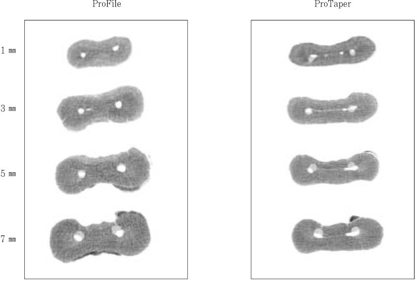
Figure 1 Examples of superimposed images. Dark gray: Cross section of root after canal shaping Light gray: Cross section of root before canal shaping
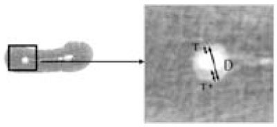
Figure 2 The drawing representing superimposed image of pre-and post-instrumentation canal shapes for measurement of centering ratio. T': Maximum extent of canal movement in one direction T: Movement in the opposite direction D: Diameter of the final canal preparation
Reference
-
1. Schilder H. Cleaning and shaping the root canal. Dent Clin North Am. 1974. 18:269–296.2. Luiten DJ, Morgan LA, Baumgartner JC, Marshall JG. A comparison of four instrumentation techniques on apical canal transportation. J Endod. 1995. 21:26–32.
Article3. Weine FS, Kelly R, Lio P. The effect of preparation procedures on the original canal shape and on apical foramen shape. J Endod. 1975. 1:255–262.
Article4. Walia HM, Brantly WA, Gerstein H. An initial investigation of the bending and torsional properties of nitinol root canal files. J Endod. 1988. 14:346–351.
Article5. Gluskin AH, Brown DC, Buchanan LS. A reconstructed computerized tomographic comparison of Ni-Ti rotary GT files versus traditional instruments in canals shaped by novice operators. Int Endod J. 2001. 34:476–484.
Article6. Taşdemir T, Aydemir UI, Inan U, Unal O. Canal preparation with Hero 642 rotary Ni-Ti instruments compared with stainless steel hand K-file assessed using computed tomography. Int Endod J. 2005. 38:402–408.
Article7. Schneider SW. A comparison of canal preparations in straight and curved canals. Oral Surg Oral Med Oral Pathol. 1971. 32:271–275.8. Calhoun G, Montgomery S. The effects of four instrumentation techniques on root canal shape. J Endod. 1988. 14:273–277.
Article9. Bramante CM, Berbert A, Borges RP. A methodology for evaluation of root canal instrumentation. J Endod. 1987. 13:243–245.
Article10. Rhodes JS, Pitt Ford TR, Lynch JA, Liepins PJ, Curtis RV. A comparison of two nickel-titanium instrumentation techniques in teeth using microcomputed tomography. Int Endod J. 2000. 33:279–285.
Article11. Peters OA, Laib A, Rüegsegger P, Barbakow F. Three-dimensional analysis of root canal geometry using high-resolution computed tomography. J Dent Res. 2000. 79:1405–1409.
Article12. Yun HH, Kim SK. A comparison of the shaping abilities of 4 nickel-titanium rotary instruments in simulated root canals. Oral Surg Oral Med Oral Pathol Oral Radiol Endod. 2003. 95:228–233.
Article13. Bergmans L, Van Cleynenbreugel J, Beullens M, Wevers M, Van Meerbeek B, Lambrechts P. Progressive versus constant tapered shaft design using NiTi rotary instruments. Int Endod J. 2003. 36:288–295.
Article14. Peters OA, Peters CI, Schönenberger K, Barbakow F. ProTaper rotary root canal preparation: effects of canal anatomy on final shape analyzed by micro CT. Int Endod J. 2003. 36:86–92.
Article15. Bergmans L, Van Cleynenbreugel J, Wevers M, Lambrechts P. Mechanical root canal preparation with Ni-Ti rotary instruments: rationale, performance and safety. Status report for the American Journal of Dentistry. Am J Dent. 2001. 14:324–333.16. Wildey WL, Senia S, Montgomery S. Another look at root canal instrumentation. Oral Surg Oral Med Oral Pathol. 1992. 74:499–507.
Article17. Ruddle CJ. Cohen S, Burns RC, editors. Cleaning and shaping the root canal system. Pathways of the Pulp. 2002. 8th ed. St. Louis, MO: Mosby;256.18. Ruddle CJ. The ProTaper Endodontic System: Geometrics, Features, and Guidelines for Use. Dent Today. 2001. 20:60–67.19. Kum KY, Spångberg L, Cha BY, Jung IY, Lee SJ, Lee CY. Shaping ability of three ProFile rotary instrumentation techniques in simulated resin root canals. J Endod. 2000. 26:719–723.
Article20. Park H. A comparison of greater taper files, ProFiles, and stainless steel files to shape curved root canals. Oral Surg Oral Med Oral Pathol Oral Radiol Endod. 2001. 91:715–718.
Article21. Schäfer E, Florek H. Efficiency of rotary nickel-titanium K3 instruments compared with stainless steel hand K-Flexofile. Part 1. Shaping ability in simulated curved canals. Int Endod J. 2003. 36:199–207.
Article22. Stone R, Zuolo M, Walton R. Apical transportaion: steel vs NiTi hand vs NiTi rotary. J Endod. 1995. 21:216. (abstract).23. Esposito PT, Cunningham CJ. A comparison of canal preparation with nickel-titanium and stainless steel instruments. J Endod. 1995. 21:173–176.
Article24. Bryant ST, Dummer PMH, Pitoni C, Bourba M, Moghal S. Shaping ability of .04 and .06 taper ProFile rotary nickel-titanium instruments in simulated root canals. Int Endod J. 1999. 32:155–164.
Article25. Peters OA, Schönenberger K, Laib A. Effects of four Ni-Ti preparation techniques on root canal geometry assessed by micro computed tomography. Int Endod J. 2001. 34:221–230.
Article
- Full Text Links
-
- Actions
-
Cited
- CITED
-
- Close
- Share
-
- Similar articles
-
- Micro-computed tomographic evaluation of a new system for root canal filling using calcium silicatebased root canal sealers
- Root canal volume change and transportation by Vortex Blue, ProTaper Next, and ProTaper Universal in curved root canals
- Relative efficacy of three Ni-Ti file systems used by undergraduates
- The instrument-centering ability of four Nickel-Titanium instruments in simulated curved root canals
- Evaluation of mesial root canal configuration of mandibular first molars using micro-computed tomography
