Imaging Sci Dent.
2014 Dec;44(4):279-285. 10.5624/isd.2014.44.4.279.
Assessment of CT numbers in limited and medium field-of-view scans taken using Accuitomo 170 and Veraviewepocs 3De cone-beam computed tomography scanners
- Affiliations
-
- 1Department of Oral Diagnosis, Piracicaba Dental School, State University of Campinas, Campinas, SP, Brazil.
- 2Department of Oral Diagnosis and Surgery, Araraquara Dental School, Sao Paulo State University, Araraquara, SP, Brazil.
- 3Section of Oral and Maxillofacial Radiology, School of Dentistry, University of California, Los Angeles, CA, USA. smallya@ucla.edu
- KMID: 1974496
- DOI: http://doi.org/10.5624/isd.2014.44.4.279
Abstract
- PURPOSE
To assess the influence of anatomic location on the relationship between computed tomography (CT) number and X-ray attenuation in limited and medium field-of-view (FOV) scans.
MATERIALS AND METHODS
Tubes containing solutions with different concentrations of K2HPO4 were placed in the tooth sockets of a human head phantom. Cone-beam computed tomography (CBCT) scans were acquired, and CT numbers of the K2HPO4 solutions were measured. The relationship between CT number and K2HPO4 concentration was examined by linear regression analyses. Then, the variation in CT number according to anatomic location was examined.
RESULTS
The relationship between K2HPO4 concentration and CT number was strongly linear. The slopes of the linear regressions for the limited FOVs were almost 2-fold lower than those for the medium FOVs. The absolute CT number differed between imaging protocols and anatomic locations.
CONCLUSION
There is a strong linear relationship between X-ray attenuation and CT number. The specific imaging protocol and anatomic location of the object strongly influence this relationship.
Keyword
MeSH Terms
Figure
-
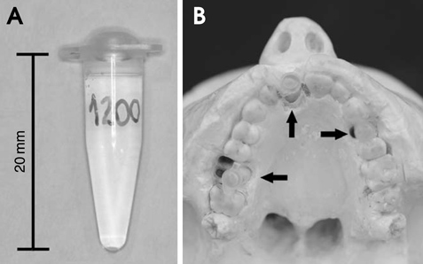
Fig. 1 A. A polypropylene tube containing K2HPO4 solution is used to attenuate X-radiation. B. K2HPO4-containing tubes (arrows) are placed in the sockets of the maxilla of a human skull phantom.
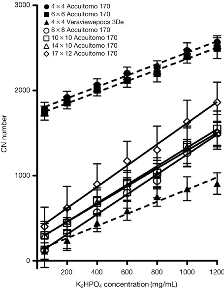
Fig. 2 A graph shows the relationship between CT number and X-ray attenuation for the limited and medium field-of-view conebeam computed tomography scans. Each symbol represents the mean±standard deviation.
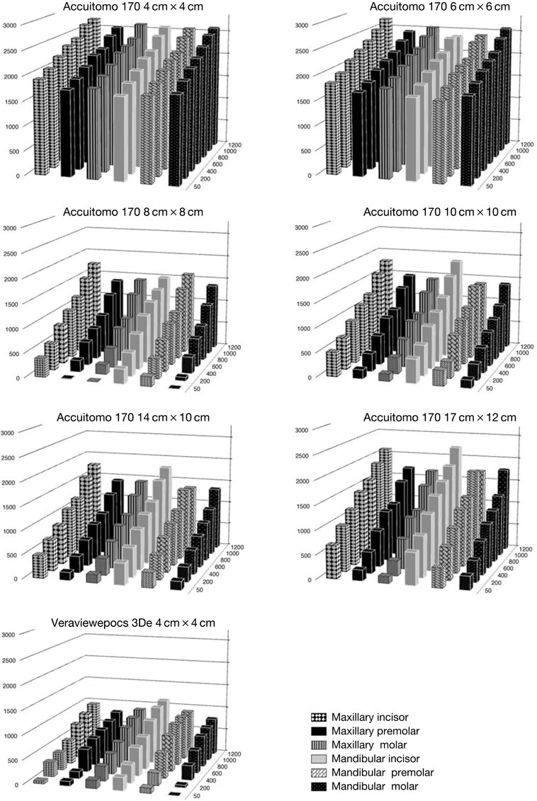
Fig. 3 A bar graph reveals the effect of anatomic location (x-axis) on the CT number (y-axis) and K2HPO4 concentration (mg/mL, z-axis) for different fields of view of the Accuitomo 170 and Veraviewepocs 3De units.
Reference
-
1. Molteni R. Prospects and challenges of rendering tissue density in Hounsfield units for cone beam computed tomography. Oral Surg Oral Med Oral Pathol Oral Radiol. 2013; 116:105–119.
Article2. Adams JE. Quantitative computed tomography. Eur J Radiol. 2009; 71:415–424.
Article3. Oliveira ML, Pedrosa EF, Cruz AD, Haiter-Neto F, Paula FJ, Watanabe PC. Relationship between bone mineral density and trabecular bone pattern in postmenopausal osteoporotic Brazilian women. Clin Oral Investig. 2013; 17:1847–1853.
Article4. Mah P, Reeves TE, McDavid WD. Deriving Hounsfield units using grey levels in cone beam computed tomography. Dentomaxillofac Radiol. 2010; 39:323–335.
Article5. Parsa A, Ibrahim N, Hassan B, Motroni A, van der, Wismeijer D. Reliability of voxel gray values in cone beam computed tomography for preoperative implant planning assessment. Int J Oral Maxillofac Implants. 2012; 27:1438–1442.6. Pauwels R, Nackaerts O, Bellaiche N, Stamatakis H, Tsiklakis K, Walker A, et al. Variability of dental cone beam CT grey values for density estimations. Br J Radiol. 2013; 86:20120135.
Article7. Valiyaparambil JV, Yamany I, Ortiz D, Shafer DM, Pendrys D, Freilich M, et al. Bone quality evaluation: comparison of cone beam computed tomography and subjective surgical assessment. Int J Oral Maxillofac Implants. 2012; 27:1271–1277.8. Lagravere MO, Carey J, Ben-Zvi M, Packota GV, Major PW. Effect of object location on the density measurement and Hounsfield conversion in a NewTom 3G cone beam computed tomography unit. Dentomaxillofac Radiol. 2008; 37:305–308.9. Oliveira ML, Tosoni GM, Lindsey DH, Mendoza K, Tetradis S, Mallya SM. Influence of anatomical location on CT numbers in cone beam computed tomography. Oral Surg Oral Med Oral Pathol Oral Radiol. 2013; 115:558–564.
Article10. Sanada S, Kawahara K, Yamamoto T, Takashima T. New tissue substitutes representing cortical bone and adipose tissue in quantitative radiology. Phys Med Biol. 1999; 44:N107–N112.
Article11. Rosset A, Spadola L, Ratib O. OsiriX: an open-source software for navigating in multidimensional DICOM images. J Digit Imaging. 2004; 17:205–216.
Article12. Silva IM, Freitas DQ, Ambrosano GM, Bóscolo FN, Almeida SM. Bone density: comparative evaluation of Hounsfield units in multislice and cone-beam computed tomography. Braz Oral Res. 2012; 26:550–556.
Article13. Naitoh M, Hirukawa A, Katsumata A, Ariji E. Evaluation of voxel values in mandibular cancellous bone: relationship between cone-beam computed tomography and multislice helical computed tomography. Clin Oral Implants Res. 2009; 20:503–506.
Article14. Nomura Y, Watanabe H, Honda E, Kurabayashi T. Reliability of voxel values from cone-beam computed tomography for dental use in evaluating bone mineral density. Clin Oral Implants Res. 2010; 21:558–562.
Article15. Parsa A, Ibrahim N, Hassan B, Motroni A, van der, Wismeijer D. Influence of cone beam CT scanning parameters on grey value measurements at an implant site. Dentomaxillofac Radiol. 2013; 42:79884780.
Article16. Cassetta M, Stefanelli LV, Pacifici A, Pacifici L, Barbato E. How accurate is CBCT in measuring bone density? A comparative CBCT-CT in vitro study. Clin Implant Dent Relat Res. 2014; 16:471–478.
Article17. Sisniega A, Zbijewski W, Badal A, Kyprianou IS, Stayman JW, Vaquero JJ, et al. Monte Carlo study of the effects of system geometry and antiscatter grids on cone-beam CT scatter distributions. Med Phys. 2013; 40:051915.
Article18. Katsumata A, Hirukawa A, Okumura S, Naitoh M, Fujishita M, Ariji E, et al. Relationship between density variability and imaging volume size in cone-beam computerized tomographic scanning of the maxillofacial region: an in vitro study. Oral Surg Oral Med Oral Pathol Oral Radiol Endod. 2009; 107:420–425.
Article
- Full Text Links
-
- Actions
-
Cited
- CITED
-
- Close
- Share
-
- Similar articles
-
- Comparison of limited- and large-volume cone-beam computed tomography using a small voxel size for detecting isthmuses in mandibular molars
- Three-dimensional imaging modalities in endodontics
- Patient radiation dose and protection from cone-beam computed tomography
- Assessment of the relationship between the mandibular third molar and the mandibular canal using panoramic radiograph and cone beam computed tomography
- Detection of maxillary second molar with two palatal roots using cone beam computed tomography: a case report
