Imaging Sci Dent.
2013 Jun;43(2):77-84. 10.5624/isd.2013.43.2.77.
Effective dose from direct and indirect digital panoramic units
- Affiliations
-
- 1Department of Oral and Maxillofacial Radiology, School of Dentistry, Oral Biology Research Institute, Chosun University, Gwangju, Korea. jdakim@chosun.ac.kr
- KMID: 1974433
- DOI: http://doi.org/10.5624/isd.2013.43.2.77
Abstract
- PURPOSE
This study aimed to provide comparative measurements of the effective dose from direct and indirect digital panoramic units according to phantoms and exposure parameters.
MATERIALS AND METHODS
Dose measurements were carried out using a head phantom representing an average man (175 cm tall, 73.5 kg male) and a limbless whole body phantom representing an average woman (155 cm tall, 50 kg female). Lithium fluoride thermoluminescent dosimeter (TLD) chips were used for the dosimeter. Two direct and 2 indirect digital panoramic units were evaluated in this study. Effective doses were derived using 2007 International Commission on Radiological Protection (ICRP) recommendations.
RESULTS
The effective doses of the 4 digital panoramic units ranged between 8.9 microSv and 37.8 microSv. By using the head phantom, the effective doses from the direct digital panoramic units (37.8 microSv, 27.6 microSv) were higher than those from the indirect units (8.9 microSv, 15.9 microSv). The same panoramic unit showed the difference in effective doses according to the gender of the phantom, numbers and locations of TLDs, and kVp.
CONCLUSION
To reasonably assess the radiation risk from various dental radiographic units, the effective doses should be obtained with the same numbers and locations of TLDs, and with standard hospital exposure. After that, it is necessary to survey the effective doses from various dental radiographic units according to the gender with the corresponding phantom.
MeSH Terms
Figure
-
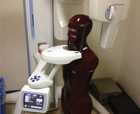
Fig. 1 The limbless female whole body phantom.
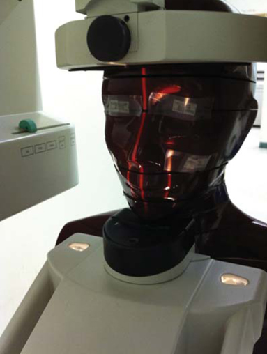
Fig. 2 The head phantom representing a man.
Reference
-
1. Rushton VE, Horner K, Worthington HV. Aspects of panoramic radiography in general dental practice. Br Dent J. 1999. 186:342–344.
Article2. Tugnait A, Clerehugh V, Hirschmann PN. Radiographic equipment and techniques used in general dental practice: a survey of general dental practitioners in England and Wales. J Dent. 2003. 31:197–203.
Article3. Hildebolt CF, Couture RA, Whiting BR. Dental photostimulable phosphor radiography. Dent Clin North Am. 2000. 44:273–297.4. Macdonald R. Digital imaging for dentists. Aust Dent J. 2001. 46:301–305.
Article5. Brennan J. An introduction to digital radiography in dentistry. J Orthod. 2002. 29:66–69.
Article6. Parks ET, Williamson GF. Digital radiography: an overview. J Contemp Dent Pract. 2002. 3:23–39.
Article7. Gijbels F, Jacobs R, Bogaerts R, Debaveye D, Verlinden S, Sanderink G. Dosimetry of digital panoramic imaging. Part I: Patient exposure. Dentomaxillofac Radiol. 2005. 34:145–149.
Article8. Ludlow JB, Ivanovic M. Comparative dosimetry of dental CBCT devices and 64-slice CT for oral and maxillofacial radiology. Oral Surg Oral Med Oral Pathol Oral Radiol Endod. 2008. 106:106–114.
Article9. Radiation protection. Protection from potential exposures: application to selected radiation sources. A report of a task group of the International Commission on Radiation Protection. Ann ICRP. 1997. 27:1–61.10. Cho JY, Han WJ, Kim EK. Absorbed and effective dose from periapical radiography by portable intraoral X-ray machine. Korean J Oral Maxillofac Radiol. 2007. 37:149–156.11. White SC. 1992 assessment of radiation risk from dental radiography. Dentomaxillofac Radiol. 1992. 21:118–126.
Article12. The 2007 Recommendations of the International Commission on Radiological Protection. ICRP Publication 103. Ann ICRP. 2007. 37:1–332.13. 1990 Recommendations of the International Commission on Radiological Protection. Ann ICRP. 1991. 21:1–201.14. Danforth RA, Clark DE. Effective dose from radiation absorbed during a panoramic examination with a new generation machine. Oral Surg Oral Med Oral Pathol Oral Radiol Endod. 2000. 89:236–243.
Article15. Lecomber AR, Downes SL, Mokhtari M, Faulkner K. Optimisation of patient doses in programmable dental panoramic radiography. Dentomaxillofac Radiol. 2000. 29:107–112.
Article16. Choi SC, Lee SM. The absorbed dose from each exposure program of the Orthopos® panoramic machine. Korean J Oral Maxillofac Radiol. 2001. 31:215–219.17. Lecomber AR, Yoneyama Y, Lovelock DJ, Hosoi T, Adams AM. Comparison of patient dose from imaging protocols for dental implant planning using conventional radiography and computed tomography. Dentomaxillofac Radiol. 2001. 30:255–259.
Article18. Ludlow JB, Davies-Ludlow LE, Brooks SL. Dosimetry of two extraoral direct digital imaging devices: NewTom cone beam CT and Orthophos Plus DS panoramic unit. Dentomaxillofac Radiol. 2003. 32:229–234.
Article19. Lee JN, Han WJ, Kim EK. Absorbed and effective dose from newly developed cone beam computed tomography in Korea. Korean J Oral Maxillofac Radiol. 2007. 37:93–102.
- Full Text Links
-
- Actions
-
Cited
- CITED
-
- Close
- Share
-
- Similar articles
-
- Absorbed and effective dose in direct and indirect digital panoramic radiography
- A study of the mandibular canal in digital panoramic radiographic images of a selected Korean population
- Combined use of direct and indirect digital impression in temporary denture fabrication
- Comparison Study of Image Quality of Direct and Indirect Conversion Digital Mammography System
- Comparative study of digital and conventional radiography for the diagnostic ability of artificial proximal surface caries
