Imaging Sci Dent.
2012 Jun;42(2):65-70. 10.5624/isd.2012.42.2.65.
Comparison of effective dose for imaging of mandible between multi-detector CT and cone-beam CT
- Affiliations
-
- 1Department of Oral and Maxillofacial Radiology and Dental Research Institute, School of Dentistry, Seoul National University, Seoul, Korea. hmslsh@snu.ac.kr
- 2Department of Oral and Maxillofacial Radiology, Dental Research Institute, and BK21 Craniomaxillofacial Life Science, School of Dentistry, Seoul National University, Seoul, Korea.
- KMID: 1974410
- DOI: http://doi.org/10.5624/isd.2012.42.2.65
Abstract
- PURPOSE
The aim of this study was to compare the effective dose for imaging of mandible between multi-detector computed tomography (MDCT) and cone-beam computed tomography (CBCT). An MDCT with low dose technique was also compared with them.
MATERIALS AND METHODS
Thermoluminescent dosimeter (TLD) chips were placed at 25 organ sites of an anthropomorphic phantom. The mandible of the phantom was exposed using 2 different types of MDCT units (Somatom Sensation 10 for standard-dose MDCT, Somatom Emotion 6 for low-dose MDCT) and 3 different CBCT units (AZ3000CT, Implagraphy, and Kavo 3D eXaM). The radiation absorbed dose was measured and the effective dose was calculated according to the ICRP 2007 report.
RESULTS
The effective dose was the highest for Somatom Sensation 10 (425.84 microSv), followed by AZ3000CT (332.4 microSv), Somatom Emotion 6 (199.38 microSv), and 3D eXaM (111.6 microSv); it was the lowest for Implagraphy (83.09 microSv). The CBCT showed significant variation in dose level with different device.
CONCLUSION
The effective doses of MDCTs were not significantly different from those of CBCTs for imaging of mandible. The effective dose of MDCT could be markedly decreased by using the low-dose technique.
Keyword
MeSH Terms
Figure
-
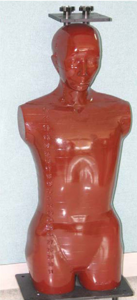
Fig. 1 Alderson radiation therapy phantom.

Fig. 2 Thermoluminescent dosimeter (TLD) chips are inserted into each of the organ structures.
Cited by 1 articles
-
Conversion coefficients for the estimation of effective dose in cone-beam CT
Dong-Soo Kim, Oyuntugs Rashsuren, Eun-Kyung Kim
Imaging Sci Dent. 2014;44(1):21-29. doi: 10.5624/isd.2014.44.1.21.
Reference
-
1. Nakagawa Y, Kobayashi K, Ishii H, Mishima A, Asada K, Ishibashi K. Preoperative application of limited cone beam computerized tomography as an assessment tool before minor oral surgery. Int J Oral Maxillofac Surg. 2002. 31:322–326.
Article2. Hounsfield GN. Computerized transverse axial scanning (tomography). 1. Description of system. Br J Radiol. 1973. 46:1016–1022.3. Ludlow JB, Davies-Ludlow LE, Brooks SL. Dosimetry of two extraoral direct digital imaging devices: NewTom cone beam CT and Orthophos Plus DS panoramic unit. Dentomaxillofac Radiol. 2003. 32:229–234.
Article4. Ludlow JB, Davies-Ludlow LE, Brooks SL, Howerton WB. Dosimetry of 3 CBCT devices for oral and maxillofacial radiology: CB Mercuray, NewTom 3G and i-CAT. Dentomaxillofac Radiol. 2006. 35:219–226.
Article5. Lee JN, Han WJ, Kim EK. Absorbed and effective dose from newly developed cone beam computed tomography in Korea. Korean J Oral Maxillofac Radiol. 2007. 37:93–102.6. Chau AC, Fung K. Comparison of radiation dose for implant imaging using conventional spiral tomography, computed tomography, and cone-beam computed tomography. Oral Surg Oral Med Oral Pathol Oral Radiol Endod. 2009. 107:559–565.
Article7. Dula K, Mini R, van der Stelt PF, Sanderink GC, Schneeberger P, Buser D. Comparative dose measurements by spiral tomography for preimplant diagnosis: the Scanora machine versus the Cranex Tome radiography unit. Oral Surg Oral Med Oral Pathol Oral Radiol Endod. 2001. 91:735–742.
Article8. Hirsch E, Wolf U, Heinicke F, Silva MA. Dosimetry of the cone beam computed tomography Veraviewepocs 3D compared with the 3D Accuitomo in different fields of view. Dentomaxillofac Radiol. 2008. 37:268–273.
Article9. Carrafiello G, Dizonno M, Colli V, Strocchi S, Pozzi Taubert S, Leonardi A, et al. Comparative study of jaws with multislice computed tomography and cone-beam computed tomography. Radiol Med. 2010. 115:600–611.
Article10. Suomalainen A, Kiljunen T, Kaser Y, Peltola J, Kortesniemi M. Dosimetry and image quality of four dental cone beam computed tomography scanners compared with multislice computed tomography scanners. Dentomaxillofac Radiol. 2009. 38:367–378.
Article11. Kim SY, Han JW, Park IW. Comparison of cone beam CT and conventional CT in absorned and effective dose. Korean J Oral Maxillofac Radiol. 2008. 38:7–15.12. Tack D, Widelec J, De Maertelaer V, Bailly JM, Delcour C, Gevenois PA. Comparison between low-dose and standard-dose multidetector CT in patients with suspected chronic sinusitis. AJR Am J Roentgenol. 2003. 181:939–944.
Article13. Groves AM, Owen KE, Courtney HM, Yates SJ, Goldstone KE, Blake GM, et al. 16-detector multislice CT: dosimetry estimation by TLD measurement compared with Monte Carlo simulation. Br J Radiol. 2004. 77:662–665.
Article14. Avendanio B, Frederiksen NL, Benson BW, Sokolowski TW. Effective dose and risk assessment from detailed narrow beam radiography. Oral Surg Oral Med Oral Pathol Oral Radiol Endod. 1996. 82:713–719.
Article15. Frederiksen NL, Benson BW, Sokolowski TW. Effective dose and risk assessment from computed tomography of the maxillofacial complex. Dentomaxillofac Radiol. 1995. 24:55–58.
Article16. Frederiksen NL, Benson BW, Sokolowski TW. Effective dose and risk assessment from film tomography used for dental implant diagnostics. Dentomaxillofac Radiol. 1994. 23:123–127.
Article17. Underhill TE, Chilvarquer I, Kimura K, Langlais RP, McDavid WD, Preece JW, et al. Radiobiologic risk estimation from dental radiology. Part I. Absorbed doses to critical organs. Oral Surg Oral Med Oral Pathol. 1988. 66:111–120.18. Mozzo P, Procacci C, Tacconi A, Martini PT, Andreis IA. A new volumetric CT machine for dental imaging based on the cone-beam technique: preliminary results. Eur Radiol. 1998. 8:1558–1564.
Article19. Loubele M, Bogaerts R, Van Dijck E, Pauwels R, Vanheusden S, Suetens P, et al. Comparison between effective radiation dose of CBCT and MSCT scanners for dentomaxillofacial applications. Eur J Radiol. 2009. 71:461–468.
Article20. Schulze D, Heiland M, Thurmann H, Adam G. Radiation exposure during midfacial imaging using 4- and 16-slice computed tomography, cone beam computed tomography systems and conventional radiography. Dentomaxillofac Radiol. 2004. 33:83–86.
Article21. Ludlow JB, Ivanovic M. Comparative dosimetry of dental CBCT devices and 64-slice CT for oral and maxillofacial radiology. Oral Surg Oral Med Oral Pathol Oral Radiol Endod. 2008. 106:106–114.
Article22. Liang X, Jacobs R, Hassan B, Li L, Pauwels R, Corpas L, et al. A comparative evaluation of Cone Beam Computed Tomography (CBCT) and Multi-Slice CT (MSCT) Part I. On subjective image quality. Eur J Radiol. 2010. 75:265–269.23. Mulkens TH, Broers C, Fieuws S, Termote JL, Bellnick P. Comparison of effective doses for low-dose MDCT and radiographic examination of sinuses in children. AJR Am J Roentgenol. 2005. 184:1611–1618.
Article24. Vassileva J, Stoyanov D. Quality control and patient dosimetry in dental cone beam CT. Radiat Prot Dosimetry. 2010. 139:310–312.
Article
- Full Text Links
-
- Actions
-
Cited
- CITED
-
- Close
- Share
-
- Similar articles
-
- Cone beam CT findings of retromolar canals: Report of cases and literature review
- Patient radiation dose and protection from cone-beam computed tomography
- Three-dimensional imaging modalities in endodontics
- Conversion coefficients for the estimation of effective dose in cone-beam CT
- Comparison of CT numbers between cone-beam CT and multi-detector CT
