Imaging Sci Dent.
2014 Mar;44(1):21-29. 10.5624/isd.2014.44.1.21.
Conversion coefficients for the estimation of effective dose in cone-beam CT
- Affiliations
-
- 1Department of Oral and Maxillofacial Radiology, Dankook University College of Dentistry, Cheonan, Korea. ekkim@dankook.ac.kr
- KMID: 1974470
- DOI: http://doi.org/10.5624/isd.2014.44.1.21
Abstract
- PURPOSE
To determine the conversion coefficients (CCs) from the dose-area product (DAP) value to effective dose in cone-beam CT.
MATERIALS AND METHODS
A CBCT scanner with four fields of view (FOV) was used. Using two exposure settings of the adult standard and low dose exposure, DAP values were measured with a DAP meter in C mode (200mm x 179 mm), P mode (154 mm x 154 mm), I mode (102 mm x 102 mm), and D mode (51 mm x 51 mm). The effective doses were also investigated at each mode using an adult male head and neck phantom and thermoluminescent chips. Linear regressive analysis of the DAP and effective dose values was used to calculate the CCs for each CBCT examination.
RESULTS
For the C mode, the P mode at the maxilla, and the P mode at the mandible, the CCs were 0.049 microSv/mGycm2, 0.067 microSv/mGycm2, and 0.064 microSv/mGycm2, respectively. For the I mode, the CCs at the maxilla and mandible were 0.076 microSv/mGycm2 and 0.095 microSv/mGycm2, respectively. For the D mode at the maxillary incisors, molars, and mandibular molars, the CCs were 0.038 microSv/mGycm2, 0.041 microSv/mGycm2, and 0.146 microSv/mGycm2, respectively.
CONCLUSION
The CCs in one CBCT device with fixed 80 kV ranged from 0.038 microSv/mGycm2 to 0.146 microSv/mGycm2 according to the imaging modes and irradiated region and were highest for the D mode at the mandibular molar.
MeSH Terms
Figure
-

Fig. 1 DIAMENTOR M4-KDK (PTW, Freiburg, Germany) used for the measurement of DAP.

Fig. 2 Adult male ART head and neck phantom (A) and TLD-100 LiF chips (B) used for the measurement of effective dose.

Fig. 3 Ionization chamber of DAP meter is located at the tube side of CBCT machine for the measurement of DAP.
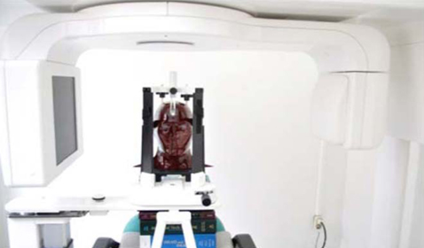
Fig. 4 Adult male ART head and neck phantom is positioned for CBCT taking.
Cited by 1 articles
-
Estimation of the effective dose of dental cone-beam computed tomography using personal computer-based Monte Carlo software
Eun-Kyung Kim, Won-Jeong Han, Jin-Woo Choi, Bulgan Battulga
Imaging Sci Dent. 2018;48(1):21-30. doi: 10.5624/isd.2018.48.1.21.
Reference
-
1. Arai Y, Tammisalo E, Iwai K, Hashimoto K, Shinoda K. Development of a compact computed tomographic apparatus for dental use. Dentomaxillofac Radiol. 1999; 28:245–248.
Article2. Momin MA, Matsumoto K, Ejima K, Asaumi R, Kawai T, Arai Y, et al. Correlation of mandibular impacted tooth and bone morphology determined by cone beam computed topography on a premise of third molar operation. Surg Radiol Anat. 2013; 35:311–318.
Article3. Patel S, Dawood A, Whaites E, Pitt Ford T. New dimensions in endodontic imaging: part 1. Conventional and alternative radiographic systems. Int Endod J. 2009; 42:447–462.
Article4. Silva MA, Wolf U, Heinicke F, Bumann A, Visser H, Hirsch E. Cone-beam computed tomography for routine orthodontic treatment planning: a radiation dose evaluation. Am J Orthod Dentofacial Orthop. 2008; 133:640.e1–640.e5.
Article5. Lofthag-Hansen S, Gröndahl K, Ekestubbe A. Cone-beam CT for preoperative implant planning in the posterior mandible: visibility of anatomic landmarks. Clin Implant Dent Relat Res. 2009; 11:246–255.
Article6. Loubele M, Bogaerts R, Van Dijck E, Pauwels R, Vanheusden S, Suetens P, et al. Comparison between effective radiation dose of CBCT and MSCT scanners for dentomaxillofacial applications. Eur J Radiol. 2009; 71:461–468.
Article7. Jeong DK, Lee SC, Huh KH, Yi WJ, Heo MS, Lee SS, et al. Comparison of effective dose for imaging of mandible between multi-detector CT and cone-beam CT. Imaging Sci Dent. 2012; 42:65–70.
Article8. Roberts JA, Drage NA, Davies J, Thomas DW. Effective dose from cone beam CT examinations in dentistry. Br J Radiol. 2009; 82:35–40.
Article9. Pauwels R, Beinsberger J, Collaert B, Theodorakou C, Rogers J, Walker A, et al. Effective dose range for dental cone beam computed tomography scanners. Eur J Radiol. 2012; 81:267–271.
Article10. Qu XM, Li G, Ludlow JB, Zhang ZY, Ma XC. Effective radiation dose of ProMax 3D cone-beam computerized tomography scanner with different dental protocols. Oral Surg Oral Med Oral Pathol Oral Radiol Endod. 2010; 110:770–776.
Article11. Davies J, Johnson B, Drage N. Effective doses from cone beam CT investigation of the jaws. Dentomaxillofac Radiol. 2012; 41:30–36.
Article12. Theodorakou C, Walker A, Horner K, Pauwels R, Bogaerts R, Jacobs R, et al. Estimation of paediatric organ and effective doses from dental cone beam CT using anthropomorphic phantoms. Br J Radiol. 2012; 85:153–160.
Article13. ICRP. Diagnostic reference levels in medical imaging: review and additional advice. A web module produced by Committee 3 of the International Commission on Radiological Protection (ICRP) [Internet]. Otawa: ICRP;2001. cited 2012 September 30. Available from: http://www.icrp.org/docs/DRL_for_web.pdf.14. Kim EK. Development of diagnostic reference level in dental x-ray examination in Korea [Internet]. Osong: Korea Food and Drug Administration;2009. cited 2012 September 30. Available from: http://rnd.mfds.go.kr.15. Goo HW. Development of the diagnostic reference level of pediatric chest radiography [Internet]. Osong: Korea Food and Drug Administration;2009. cited 2012 September 30. Available from: http://rnd.mfds.go.kr.16. Kim HJ. Study for establishment of diagnostic reference level of patient dose in skull radiography [Internet]. Osong: Korea Food and Drug Administration;2009. cited 2012 September 30. Available from: http://rnd.mfds.go.kr/.17. Sung DW. Investigation of patient dose for diagnostic reference levels (DRL) in radiographic examination: National survey in Korea [Internet]. Osong: Korea Food and Drug Administration;2011. cited 2012 September 30. Available from: http://rnd.mfds.go.kr.18. Thilander-Klang A, Helmrot E. Methods of determining the effective dose in dental radiology. Radiat Prot Dosimetry. 2010; 139:306–309.
Article19. Helmrot E, Alm Carlsson G. Measurement of radiation dose in dental radiology. Radiat Prot Dosimetry. 2005; 114:168–171.
Article20. Holroyd JR, Walker A. HPA-RPD-065. Recommendations for the design of X-ray facilities and quality assurance of dental Cone Beam CT (Computed Tomography) systems. Chilton: Health Protection Agency;2010.21. Lofthag-Hansen S, Thilander-Klang A, Ekestubbe A, Helmrot E, Gröndahl K. Calculating effective dose on a cone beam computed tomography device: 3D Accuitomo and 3D Accuitomo FPD. Dentomaxillofac Radiol. 2008; 37:72–79.
Article22. Schultz FW, Zoetelief J. Dose conversion coefficients for interventional procedures. Radiat Prot Dosimetry. 2005; 117:225–230.
Article23. Compagnone G, Giampalma E, Domenichelli S, Renzulli M, Golfieri R. Calculation of conversion factors for effective dose for various interventional radiology procedures. Med Phys. 2012; 39:2491–2498.
Article24. Hart D, Wall BF. NRPB-W4. Radiation exposure of the UK population from medical and dental X-ray examinations. Chilton: National Radiological Protection Board;2002.25. Looe HK, Eenboom F, Chofor N, Pfaffenberger A, Steinhoff M, Rühmann A, et al. Conversion coefficients for the estimation of effective doses in intraoral and panoramic dental radiology from dose-area product values. Radiat Prot Dosimetry. 2008; 131:365–373.
Article26. Looe HK, Eenboom F, Chofor N, Pfaffenberger A, Sering M, Rühmann A, et al. Dose-area product measurements and determination of conversion coefficients for the estimation of effective dose in dental lateral cephalometric radiology. Radiat Prot Dosimetry. 2007; 124:181–186.
Article27. Ludlow JB, Ivanovic M. Comparative dosimetry of dental CBCT devices and 64-slice CT for oral and maxillofacial radiology. Oral Surg Oral Med Oral Pathol Oral Radiol Endod. 2008; 106:106–114.
Article28. The 2007 Recommendations of the International Commission on Radiological Protection. ICRP publication 103. Ann ICRP. 2007; 37:1–332.29. Radiation protection No. 172. Evidence based guidelines on cone beam CT for dental and maxillofacial radiology [Internet]. Luxemburg: European commission;2012. cited 2012 September 30. Available from: http://ec.europa.eu/energy/nuclear/radiation_protection/doc/publication/172.pdf.
- Full Text Links
-
- Actions
-
Cited
- CITED
-
- Close
- Share
-
- Similar articles
-
- Erratum to: Conversion coefficients for the estimation of effective dose in cone-beam CT
- Evaluation of Radiation Dose for Dual Energy CBCT Using Multi-Grid Device
- Three-dimensional imaging modalities in endodontics
- Predicting Factors for Conversion from Fluoroscopy Guided Percutaneous Transthoracic Needle Biopsy to Cone-Beam CT Guided Percutaneous Transthoracic Needle Biopsy
- Patient radiation dose and protection from cone-beam computed tomography
