Highlights from the 52nd Seminar of the Korean Society of Gastrointestinal Endoscopy
- Affiliations
-
- 1Department of Internal Medicine, Catholic University of Daegu School of Medicine, Daegu, Korea.
- 2Center for Gastric Cancer, National Cancer Center, Goyang, Korea.
- 3Department of Gastroenterology, Gachon University Gil Medical Center, Gachon University College of Medicine, Incheon, Korea.
- 4Department of Internal Medicine and Liver Research Institute, Seoul National University College of Medicine, Seoul, Korea.
- 5Department of Gastroenterology, CHA Bundang Medical Center, CHA University, Seongnam, Korea. hahmkb@cha.ac.kr
- KMID: 1964267
- DOI: http://doi.org/10.5946/ce.2015.48.4.269
Abstract
- In this July issue of Clinical Endoscopy, state-of-the-art articles selected from the lectures delivered during the 52nd Seminar of the Korean Society of Gastrointestinal Endoscopy (KSGE) on March 29, 2015 are covered, focusing on highlighted educational contents relevant to either diagnostic or therapeutic gastrointestinal (GI) endoscopy. Our society, the KSGE, has continued to host this opportunity for annual seminars twice a year over the last 26 years and it has become a large-scale prestigious seminar accommodating over 4,000 participants. Definitely, the KSGE seminar is considered as one of the premier state-of-the-art seminars dealing with GI endoscopy, appealing to both the beginner and advanced experts. Lectures, live demonstrations, hands-on courses, as well as an editor school, which was an important consensus meeting on how to upgrade our society journal, Clinical Endoscopy, to a Science Citation Index (Expanded) designation were included in this seminar. The 52nd KSGE seminar consisted of more than 20 sessions, including special lectures, concurrent sessions for GI endoscopy nurses, and sessions exploring new technologies. This is a very special omnibus article to highlight the core contents divided into four sessions: upper GI tract, lower GI tract, pancreatobiliary system, and other specialized sessions.
MeSH Terms
Figure
-
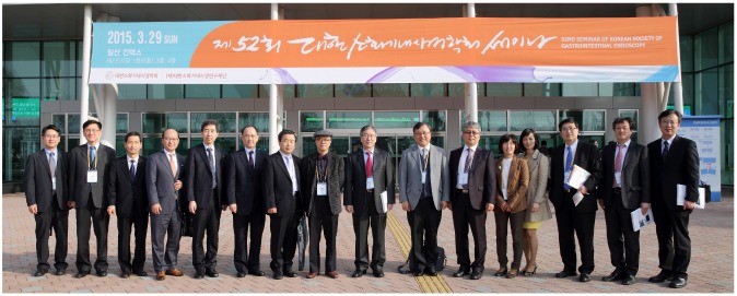
Fig. 1 A group photo showing the great contribution at the successful 52nd Seminar of the Korean Society of Gastrointestinal Endoscopy. From the left, Jae Myung Park (Seoul St. Mary's Hospital, College of Medicine, The Catholic University of Korea, Seoul), Sam Ryong Jee (Inje University Busan Paik Hospital, Inje University College of Medicine, Busan), Jeong Seop Moon (Inje University Seoul Paik Hospital, Inje University College of Medicine, Seoul), Il Kwun Chung (Soonchunhyang University Cheonan Hospital, Soonchunhyang University College of Medicine, Cheonan), Yoon Tae Jeen (Korea University Anam Hospital, Korea University College of Medicine, Seoul), Oh Young Lee (Hanyang University Seoul Hospital, Hanyang University College of Medicine, Seoul), Yong Woon Shin (Inha University Hospital, Inha University School of Medicine, Incheon), Sang Yong Seol (Inje University Busan Paik Hospital, Inje University College of Medicine, Busan), Sei Jin Youn (Chungbuk National University Hospital, Chungbuk National University College of Medicine, Cheongju), Myung-Gyu Choi (Seoul St. Mary's Hospital, College of Medicine, The Catholic University of Korea, Seoul), Chang-Hun Yang (Dongguk University Gyeongju Hospital, Dongguk University College of Medicine, Gyeongju), Eun Young Kim (Daegu Catholic University Medical Center, Catholic University of Daegu School of Medicine, Daegu), Ki-Nam Shim (Ewha Womans University Mokdong Hospital, Ewha Womans University School of Medicine, Seoul), Ki Baik Hahm (CHA Bundang Medical Center, CHA University, Seongnam), Don Haeng Lee (Inha University Hospital, Inha University School of Medicine, Incheon), and Young Seok Cho (Uijeongbu St. Mary's Hospital, College of Medicine, The Catholic University of Korea, Uijeongbu, Korea).
Reference
-
1. Park KS. Introduction to starting upper gastrointestinal endoscopy: proper insertion, complete observation, and appropriate photographing. Clin Endosc. 2015; 48:279–284.
Article2. Park WG, Shaheen NJ, Cohen J, et al. Quality indicators for EGD. Gastrointest Endosc. 2015; 81:17–30. PMID: 25480101.
Article3. Standards of Practice Committee of the American Society for Gastrointestinal Endoscopy. Lichtenstein DR, Jagannath S, et al. Sedation and anesthesia in GI endoscopy. Gastrointest Endosc. 2008; 68:815–826. PMID: 18984096.
Article4. Moon HS. Improving the endoscopic detection rate in patients with early gastric cancer. Clin Endosc. 2015; 48:291–296.
Article5. Vakil N, van Zanten SV, Kahrilas P, Dent J, Jones R. Global Consensus Group. The Montreal definition and classification of gastroesophageal reflux disease: a global evidence-based consensus. Am J Gastroenterol. 2006; 101:1900–1920. PMID: 16928254.
Article6. Lu CL. Silent gastroesophageal reflux disease. J Neurogastroenterol Motil. 2012; 18:236–238. PMID: 22837870.
Article7. Dixon MF, Genta RM, Yardley JH, Correa P. Classification and grading of gastritis. The updated Sydney System. International Workshop on the Histopathology of Gastritis, Houston 1994. Am J Surg Pathol. 1996; 20:1161–1181. PMID: 8827022.8. Cho JW. The role of endosonography in the staging of gastrointestinal cancers. Clin Endosc. 2015; 48:297–301.
Article9. Kim HU. Diagnostic and treatment approaches for refractory peptic ulcers. Clin Endosc. 2015; 48:285–290.
Article10. Hassan C, Bretthauer M, Kaminski MF, et al. Bowel preparation for colonoscopy: European Society of Gastrointestinal Endoscopy (ESGE) guideline. Endoscopy. 2013; 45:142–150. PMID: 23335011.
Article11. Johnson DA, Barkun AN, Cohen LB, et al. Optimizing adequacy of bowel cleansing for colonoscopy: recommendations from the US multi-society task force on colorectal cancer. Gastroenterology. 2014; 147:903–924. PMID: 25239068.
Article12. Hassan C, Fuccio L, Bruno M, et al. A predictive model identifies patients most likely to have inadequate bowel preparation for colonoscopy. Clin Gastroenterol Hepatol. 2012; 10:501–506. PMID: 22239959.13. Desai J, Granger CB, Weitz JI, Aisenberg J. Novel oral anticoagulants in gastroenterology practice. Gastrointest Endosc. 2013; 78:227–239. PMID: 23725876.
Article14. Rex DK, Goodwine BW. Method of colonoscopy in 42 consecutive patients presenting after prior incomplete colonoscopy. Am J Gastroenterol. 2002; 97:1148–1151. PMID: 12014719.
Article15. Almadi MA, Ghosh S, Aljebreen AM. Differentiating intestinal tuberculosis from Crohn's disease: a diagnostic challenge. Am J Gastroenterol. 2009; 104:1003–1012. PMID: 19240705.
Article16. Epstein D, Watermeyer G, Kirsch R. Review article: the diagnosis and management of Crohn's disease in populations with high-risk rates for tuberculosis. Aliment Pharmacol Ther. 2007; 25:1373–1388. PMID: 17539977.
Article17. Oba S, Tanaka S, Sano Y, Oka S, Chayama K. Current status of narrow-band imaging magnifying colonoscopy for colorectal neoplasia in Japan. Digestion. 2011; 83:167–172. PMID: 21266811.
Article18. Kim TO. Colorectal subepithelial lesions. Clin Endosc. 2015; 48:302–307.
Article19. Nishihara R, Wu K, Lochhead P, et al. Long-term colorectal-cancer incidence and mortality after lower endoscopy. N Engl J Med. 2013; 369:1095–1105. PMID: 24047059.
Article20. Pabby A, Schoen RE, Weissfeld JL, et al. Analysis of colorectal cancer occurrence during surveillance colonoscopy in the dietary Polyp Prevention Trial. Gastrointest Endosc. 2005; 61:385–391. PMID: 15758908.
Article21. Kim J. Endoscopic ultrasound-guided treatment of pancreatic cystic and solid masses. Clin Endosc. 2015; 48:308–311.
Article
- Full Text Links
-
- Actions
-
Cited
- CITED
-
- Close
- Share
-
- Similar articles
-
- Highlights from the 50th Seminar of the Korean Society of Gastrointestinal Endoscopy
- Highlights of the 48th Seminar of Korean Society of Gastrointestinal Endoscopy
- Meeting Report and Special Issue Preface: The 54th Seminar of the Korean Society of Gastrointestinal Endoscopy
- Present Status and Education of Digestive Endoscopy in Korea
- Past, Present, and Future of the Korea-Japan Joint Symposium on Gastrointestinal Endoscopy
