Highlights of the 48th Seminar of Korean Society of Gastrointestinal Endoscopy
- Affiliations
-
- 1Department of Gastroenterology, Gachon University Gil Medical Center, Gachon University of Medicine and Science, Incheon, Korea.
- 2Center for Gastric Cancer, National Cancer Center, Goyang, Korea.
- 3Department of Internal Medicine, Catholic University of Daegu School of Medicine, Daegu, Korea.
- 4Division of Gastroenterology, Department of Internal Medicine, Kyung Hee University School of Medicine, Seoul, Korea.
- 5Department of Gastroenterology, CHA Bundang Medical Center, CHA University, Seongnam, Korea. hahmkb@cha.ac.kr
- KMID: 2048921
- DOI: http://doi.org/10.5946/ce.2013.46.3.203
Abstract
- This special May issue of Clinical Endoscopy discusses the tutorial contents dealing with either the diagnostic or therapeutic gastrointestinal (GI) endoscopy that contain very fundamental and essential points in this filed. The seminar of Korean Society of Gastrointestinal Endoscopy (KSGE) had positioned as one of prime educational seminars covering the very beginner to advanced experts of GI endoscopy. Besides of four rooms allocated for each lecture, two additional rooms were open for either live demonstration or hands-on course, covering totally 20 sessions including one special lecture. Among these prestigious lectures, 12 lectures were selected for the current review articles in this special issue of Clinical Endoscopy journal. Basic course for beginner to advanced tips to expert were all covered in this seminar. This introductory review prepared by four associated editors of Clinical Endoscopy contained core contents divided into four sessions-upper gut, lower gut, pancreaticobiliary, and specialized topic session part-to enhance understandings not covered by enlisted review articles in this issue.
Keyword
MeSH Terms
Figure
-
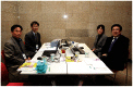
Fig. 1 The 48th Seminar of Korean Society of Gastrointestinal Endoscopy, editorial staffs for special issue, From left, Prof. Il Ju Choi (National Cancer Center), Prof. Kwang An Kwon (Gachon University Gil Medical Center), Ms. Ha Young Park, and Prof. Ki Baik Hahm (CHA Bundang Medical Center CHA University).
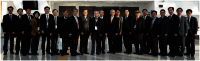
Fig. 2 The 48th Seminar of Korean Society of Gastrointestinal Endoscopy, whole members of organization committee. From left, Dae Young Cheung (Yeouido St. Mary's Hospital, The Catholic University of Korea College of Medicine), Ja Seol Koo (Korea University Ansan Hospital), Don Haeng Lee (Inha University Hospital), Eun Young Kim (Daegu Catholic University Medical Center), Young Soo Moon (Inje University Haeundae Paik Hospital), Ho Gak Kim (Daegu Catholic University Medical Center), Seok Reyol Choi (Dong-A University Hospital), Chang-Hun Yang (Dongguk University Gyeongju Hospital), Myung-Gyu Choi (Seoul St. Mary's Hospital, The Catholic University of Korea College of Medicine), Sang Yong Seol (Inje University Busan Paik Hospital), Chang Duck Kim (Korea University Anam Hospital), Young-Tae Bak (Korea University Guro Hospital), Sung Koo Lee (Asan Medical Center, University of Ulsan College of Medicine), Ki Baik Hahm (CHA Bundang Medical Center, CHA University), Il Kwun Chung (Soonchunhyang University Cheonan Hospital), Yong-Tae Kim (Seoul National University College of Medicine), Seok Ho Dong (Kyung Hee University School of Medicine), Eun Taek Park (Kosin University Gospel Hospital), Chang Hwan Park (Chonnam National University Hospital), Chang Hwan Choi (Chung-Ang University Hospital), Bo-In Lee (Incheon St. Mary's Hospital, The Catholic University of Korea College of Medicine).
Cited by 1 articles
-
Introduction to Starting Upper Gastrointestinal Endoscopy: Proper Insertion, Complete Observation, and Appropriate Photographing
Kyung Sik Park
Clin Endosc. 2015;48(4):279-284. doi: 10.5946/ce.2015.48.4.279.
Reference
-
1. Park KS. Observable laryngopharyngeal lesions during the upper gastrointestinal endoscopy. Clin Endosc. 2013; 46:224–229.
Article2. Heo J, Jeon SW. The clinical significance and management of noncurative endoscopic resection in early gastric cancer. Clin Endosc. 2013; 46:235–238.
Article3. American Society of Anesthesiologists Task Force on Sedation and Analgesia by Non-Anesthesiologists. Practice guidelines for sedation and analgesia by non-anesthesiologists. Anesthesiology. 2002; 96:1004–1017. PMID: 11964611.4. Jung Y, Lee SH. How do I overcome difficulties in insertion? Clin Endosc. 2012; 45:278–281. PMID: 22977817.
Article5. Suzuki T, Matsushima M, Tsukune Y, et al. Double-balloon endoscopy versus magnet-imaging enhanced colonoscopy for difficult colonoscopies, a randomized study. Endoscopy. 2012; 44:38–42. PMID: 22143991.
Article6. ASGE Standards of Practice Committee. Anderson MA, Ben-Menachem T, et al. Management of antithrombotic agents for endoscopic procedures. Gastrointest Endosc. 2009; 70:1060–1070. PMID: 19889407.
Article7. Vormbrock K, Mönkemüller K. Difficult colon polypectomy. World J Gastrointest Endosc. 2012; 4:269–280. PMID: 22816006.
Article8. Efthymiou M, Taylor AC, Desmond PV, Allen PB, Chen RY. Biopsy forceps is inadequate for the resection of diminutive polyps. Endoscopy. 2011; 43:312–316. PMID: 21412704.
Article9. Liu S, Ho SB, Krinsky ML. Quality of polyp resection during colonoscopy: are we achieving polyp clearance? Dig Dis Sci. 2012; 57:1786–1791. PMID: 22461018.
Article10. Pohl H, Srivastava A, Bensen SP, et al. Incomplete polyp resection during colonoscopy-results of the complete adenoma resection (CARE) study. Gastroenterology. 2013; 144:74–80. PMID: 23022496.
Article11. Rex DK, Kahi C, O'Brien M, et al. The American Society for Gastrointestinal Endoscopy PIVI (Preservation and Incorporation of Valuable Endoscopic Innovations) on real-time endoscopic assessment of the histology of diminutive colorectal polyps. Gastrointest Endosc. 2011; 73:419–422. PMID: 21353837.
Article12. Hewett DG, Kaltenbach T, Sano Y, et al. Validation of a simple classification system for endoscopic diagnosis of small colorectal polyps using narrow-band imaging. Gastroenterology. 2012; 143:599–607. PMID: 22609383.
Article13. Hamilton MJ. The valuable role of endoscopy in inflammatory bowel disease. Diagn Ther Endosc. 2012; 2012:467979. PMID: 22474403.
Article14. Jung SA. Differential diagnosis of inflammatory bowel disease: what is the role of colonoscopy? Clin Endosc. 2012; 45:254–262. PMID: 22977813.
Article15. Bourreille A, Ignjatovic A, Aabakken L, et al. Role of small-bowel endoscopy in the management of patients with inflammatory bowel disease: an international OMED-ECCO consensus. Endoscopy. 2009; 41:618–637. PMID: 19588292.
Article16. Travis SP, Schnell D, Krzeski P, et al. Developing an instrument to assess the endoscopic severity of ulcerative colitis: the Ulcerative Colitis Endoscopic Index of Severity (UCEIS). Gut. 2012; 61:535–542. PMID: 21997563.
Article17. Hassan C, Zullo A, De Francesco V, et al. Systematic review: endoscopic dilatation in Crohn's disease. Aliment Pharmacol Ther. 2007; 26:1457–1464. PMID: 17903236.
Article18. East JE. Colonoscopic cancer surveillance in inflammatory bowel disease: what's new beyond random biopsy? Clin Endosc. 2012; 45:274–277. PMID: 22977816.
Article19. Farraye FA, Odze RD, Eaden J, Itzkowitz SH. AGA technical review on the diagnosis and management of colorectal neoplasia in inflammatory bowel disease. Gastroenterology. 2010; 138:746–774. PMID: 20141809.
Article20. Lee TH. Technical tips and issues of biliary stenting, focusing on malignant hilar obstruction. Clin Endosc. 2013; 46:260–266.
Article21. Rastogi A. Optical diagnosis of small colorectal polyp histology with high-definition colonoscopy using narrow band imaging. Clin Endosc. 2013; 46:120–129. PMID: 23614121.
Article22. Kim KO, Ku YS. Is image-enhanced endoscopy useful for the diagnosis and treatment of gastrointestinal tumor? Clin Endosc. 2013; 46:248–250.
Article23. Kim EY. Introduction: value of endoscopic ultrasound-guided fine needle aspiration. Clin Endosc. 2012; 45:115–116. PMID: 22866249.
Article24. Cho JW. The Role of endoscopic ultrasonography in T staging: early gastric cancer and esophageal cancer. Clin Endosc. 2013; 46:239–242.
Article25. Hong KH, Lim YJ. Recent update of gastrointestinal endoscope reprocessing. Clin Endosc. 2013; 46:267–273.
Article26. Lee YK, Park JB. Steps of reprocessing and equipments. Clin Endosc. 2013; 46:274–279.
Article
- Full Text Links
-
- Actions
-
Cited
- CITED
-
- Close
- Share
-
- Similar articles
-
- Highlights from the 50th Seminar of the Korean Society of Gastrointestinal Endoscopy
- Highlights from the 52nd Seminar of the Korean Society of Gastrointestinal Endoscopy
- Meeting Report and Special Issue Preface: The 54th Seminar of the Korean Society of Gastrointestinal Endoscopy
- Present Status and Education of Digestive Endoscopy in Korea
- Highlights of International Digestive Endoscopy Network 2013
