Histologic Estimation of Intrauterine Retention Time after Fetal Death
- Affiliations
-
- 1Department of Medical Humanities and Social Sciences, University of Ulsan College of Medicine, Seoul, Korea. jhk@amc.seoul.kr
- KMID: 1928015
- DOI: http://doi.org/10.7580/kjlm.2013.37.4.191
Abstract
- The intrauterine retention time (IURT) after fetal death can be estimated from the loss of nuclear basophilia. We therefore attempted to derive an autolysis equation to estimate IURT in experimental rat fetuses and human fetal autopsy slides. The degree of loss of nuclear basophilia in various tissues was assessed by hematoxylin and eosin (H & E) staining. Fetal rat tissues showed different rates of autolysis, allowing for the construction of an experimental autolysis curve. We also reviewed the H & E stained slides obtained from 27 human fetal autopsy cases with well-documented death intervals. The degree of autolysis in various tissues was evaluated using percentile scores (PS). Using the findings from H&E staining, we derived the equation Ln (PS/[100-PS]) = 2.62716-0.02377 x IURT. However, this equation or autolysis scores showed some limitations. Owing to the inconsistency of PS, this equation is reliably applicable only within 24 hours of intrauterine fetal death. In the fetal autopsy review, fetal hydrops, local effusion, and sepsis also contributed to accelerated autolysis.
Keyword
MeSH Terms
Figure
-

Fig. 1. The loss of nuclear basophilia was assessed according to the Genest's criteria: a) Kidney proximal tubules (arrow, PS = 100) and distal tubules (double line arrow, PS = 100) (H & E, ×400). b) Kidney collecting tubule (arrow, PS = 50, double line arrow, PS = 0) (H & E, ×400). c) Heart cardiac myocytes show (PS = 100), (H & E, ×400). d) Cardiac myocytes (PS = 50), (H & E, ×200). e) Bronchial epithelium shows (PS = 100), (H & E, × 400). f) Bronchial epithelium shows (PS = 50), (H & E, ×400). g) Bronchial cartilage (PS = 100), (H & E, ×400). h) Bronchial chondrocytes (PS = 50), (H & E, ×400). i) Outer fetal zone of adrenal gland shows (arrow, PS = 100) and inner permanent zone (double line arrow) shows (arrow, PS = 50), (H & E, ×200). j) Hepatocytes (arrow, PS = 100) (arrow) and hematopoietic cells (double line arrow), (H & E, ×400). k) Hepatocytes (PS = 50), (H & E, ×400). l) Leukocyte common antigen staining of thymocytes shows (PS = 100), (H & E, ×400) (PS, percentile score).

Fig. 2. Autolysis curve derived from fetal rats. Percentile Score (PS) shows a steep downward slope after 24 hours immersion time (IT).
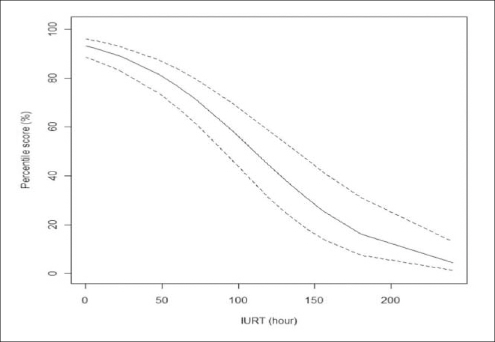
Fig. 3. IURT derived from the percentile score (PS) of H & E. Ln(PS/(100-PS)) = 2.62716 - 0.02377 × IURT, which is shown in the middle line. The other lines represent the upper and lower limits of Intrauterine Retention Time (IURT).
Reference
-
1. Shanklin DR. Fetal maceration. 2. An analysis of 53 human stillborn infants. Am J Obstet Gynecol. 1964; 88:224–9.2. Babala J. Practical significance and various new aspects arising from the pathological-anatomical analysis of intrauterine dead fetuses. Bratisl Lek Listy. 1970; 54:210–5.3. Genest DR, Williams MA, Greene MF. Estimating the time of death in stillborn fetuses: I. Histologic evaluation of fetal organs; an autopsy study of 150 stillborns. Obstet Gynecol. 1992; 80:575–84.4. Langman J. Medical embryology. Baltimore: Williams & Wilkins;1981. 351.5. Dillman RC, Dennis SM. Sequential sterile autolysis in the ovine fetus: microscopic changes. Am J Vet Res. 1979; 40:321–5.
- Full Text Links
-
- Actions
-
Cited
- CITED
-
- Close
- Share
-
- Similar articles
-
- Analysis of placental pathological findings contributing to intrauterine fetal death
- Postpartum Choriocarcinoma Preceded by Neonatal Anemia and Intrauterine Fetal Death: A case Report
- Clinical Characteristics of Intrauterine Fetal Death
- A Case Of Intrauterine Fetal Death Due To Stricture Of The Umbilical Cord
- Ectopic Umbilical Liver Associated with Intrauterine Fetal Death: An autopsy case