Visibility of Internal Target Volume of Dynamic Tumors in Free-breathing Cone-beam Computed Tomography for Image Guided Radiation Therapy
- Affiliations
-
- 1Center for Advanced Radiotherapy Technologies and Department of Radiation Medicine and Applied Sciences, University of California San Diego, La Jolla, CA, USA. wysong@ucsd.edu
- 2Department of Physics, San Diego State University, San Diego, CA, USA.
- 3Department of Electrical and Computer Engineering, University of California San Diego, La Jolla, CA, USA.
- KMID: 1910562
- DOI: http://doi.org/10.14316/pmp.2013.24.4.220
Abstract
- Respiratory-induced dynamic tumors render free-breathing cone-beam computed tomography (FBCBCT) images with motion artifacts complicating the task of quantifying the internal target volume (ITV). The purpose of this paper is to study the visibility of the revealed ITV when the imaging dose parameters, such as the kVp and mAs, are varied. The Trilogy(TM) linear accelerator with an On-Board Imaging (OBI(TM)) system was used to acquire low-imaging-dose-mode (LIDM: 110 kVp, 20 mA, 20 ms/frame) and high-imaging-dose-mode (HIDM: 125 kVp, 80 mA, 25 ms/frame) FBCBCT images of a 3-cm diameter sphere (density=0.855 g/cm3) moving in accordance to various sinusoidal breathing patterns, each with an unique inhalation-to-exhalation (I/E) ratio, amplitude, and period. In terms of image ITV contrast, there was a small overall average change of the ITV contrast when going from HIDM to LIDM of 6.5+/-5.1% for all breathing patterns. As for the ITV visible volume measurements, there was an insignificant difference between the ITV of both the LIDM- and HIDM-FBCBCT images with an average difference of 0.5+/-0.5%, for all cases, despite the large difference in the imaging dose (approximately five-fold difference of ~0.8 and 4 cGy/scan). That indicates that the ITV visibility is not very sensitive to changes in imaging dose. However, both of the FBCBCT consistently underestimated the true ITV dimensions by up to 34.8% irrespective of the imaging dose mode due to significant motion artifacts, and thus, this imaging technique is not adequate to accurately visualize the ITV for image guidance. Due to the insignificant impact of imaging dose on ITV visibility, a plausible, alternative strategy would be to acquire more X-ray projections at the LIDM setting to allow 4DCBCT imaging to better define the ITV, and at the same time, maintain a reasonable imaging dose, i.e., comparable to a single HIDM-FBCBCT scan.
Keyword
MeSH Terms
Figure
-
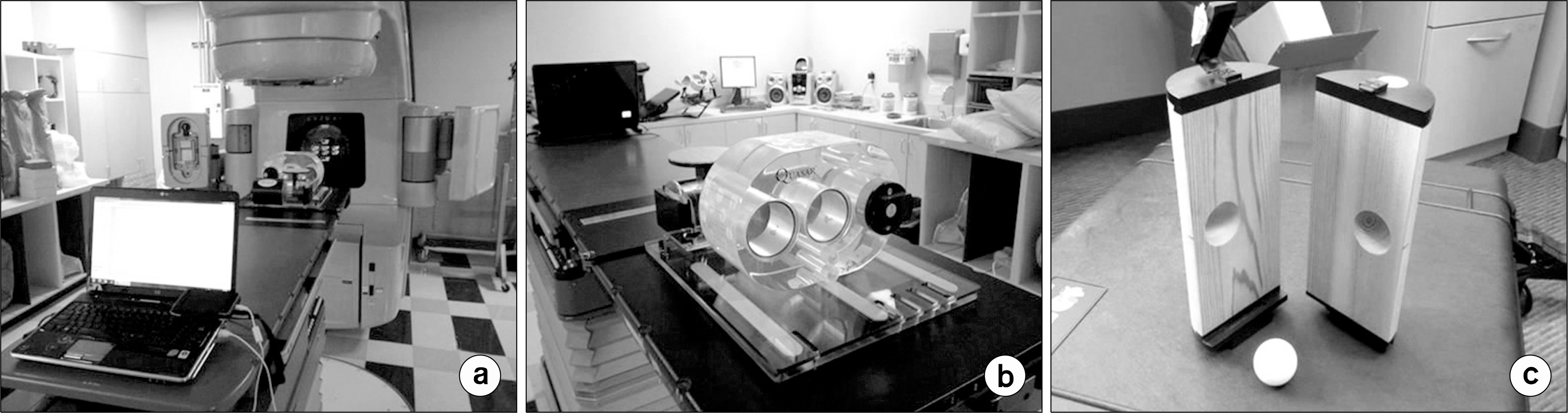
Fig. 1. “Experimental Setup”. (a) Front-side view of the setup. (b) Back-side view of the setup with the cylindrical cedar wood containing the sphere placed within the Multi-Purpose Body Phantom. (c) The cylindrical cedar wood and the sphere.
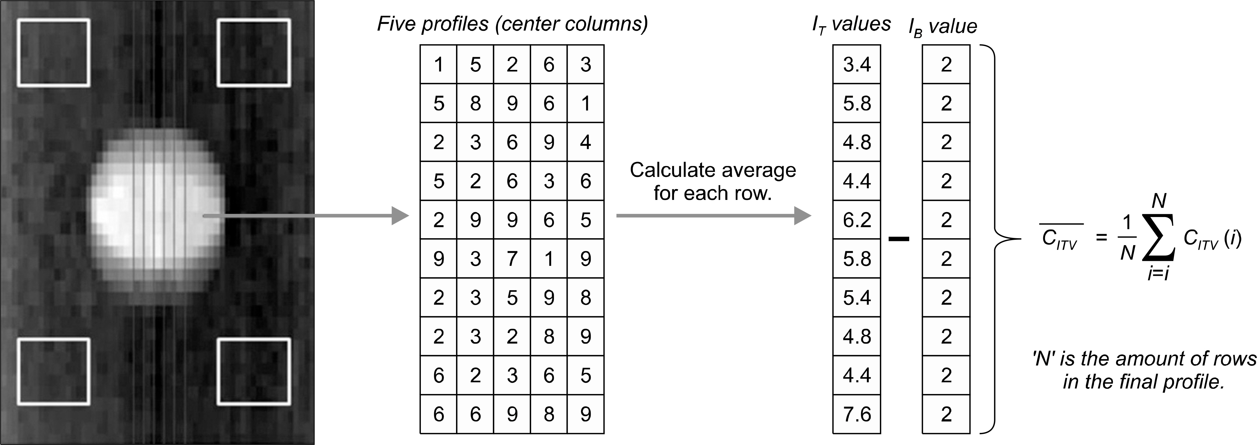
Fig. 2. “ITV Contrast Calculation”. An illistration of how the ITV contrast is calculated using an example coronal image slice of a FBCBCT scan. The four yellow squares represent the 10×10-pixels ROIs that were used to calculate the Ib. The average pixel values of the five central colums of the ITV were used to calculate the the IT.

Fig. 3. “Central Profile Comparison”. An example case showing the central profiles of the LIDM and HIDM FBCBCT images with I/E ratios ranging from 0.2632 through 1, amplitude=1 cm, and period=6 seconds.
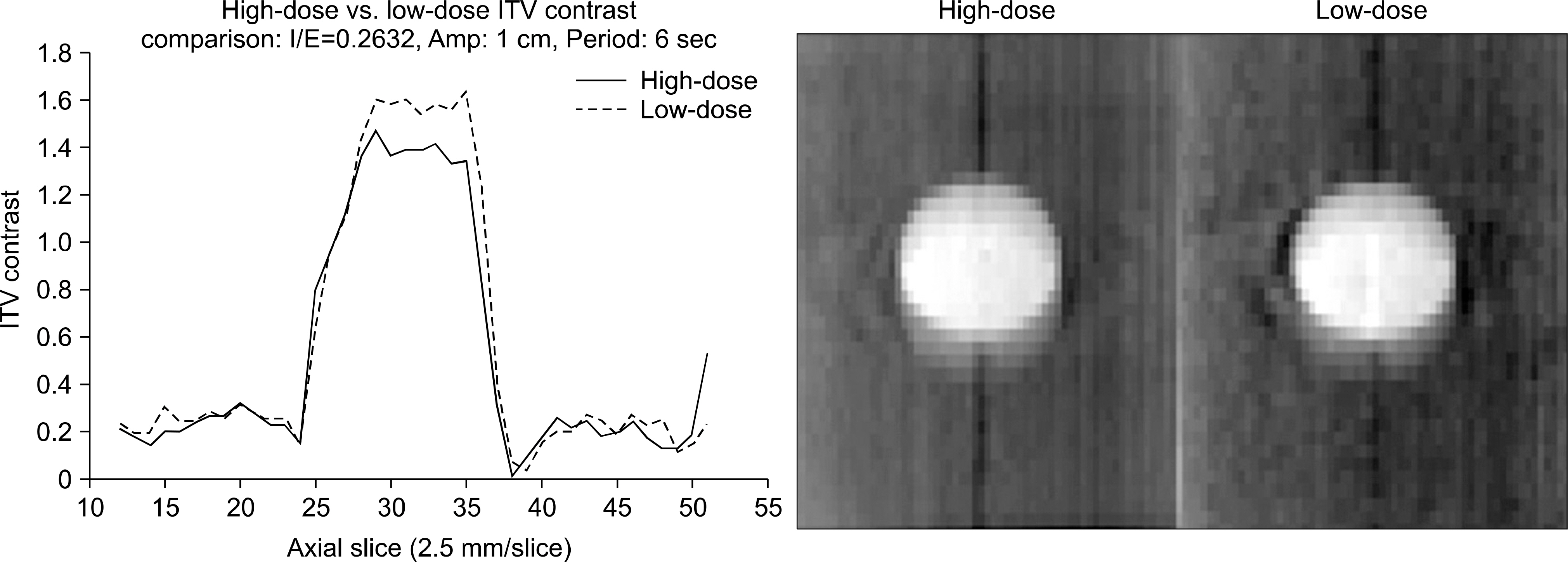
Fig. 3. Continued.

Fig. 4. “HD_LD Comparison”. As seen for both amplitudes, the accuracy of volume measurement worsens as the I/E ratio decreases. Despite that, the difference in volume measurement with HIDM and LIDM FBCBCTs is insignificant.
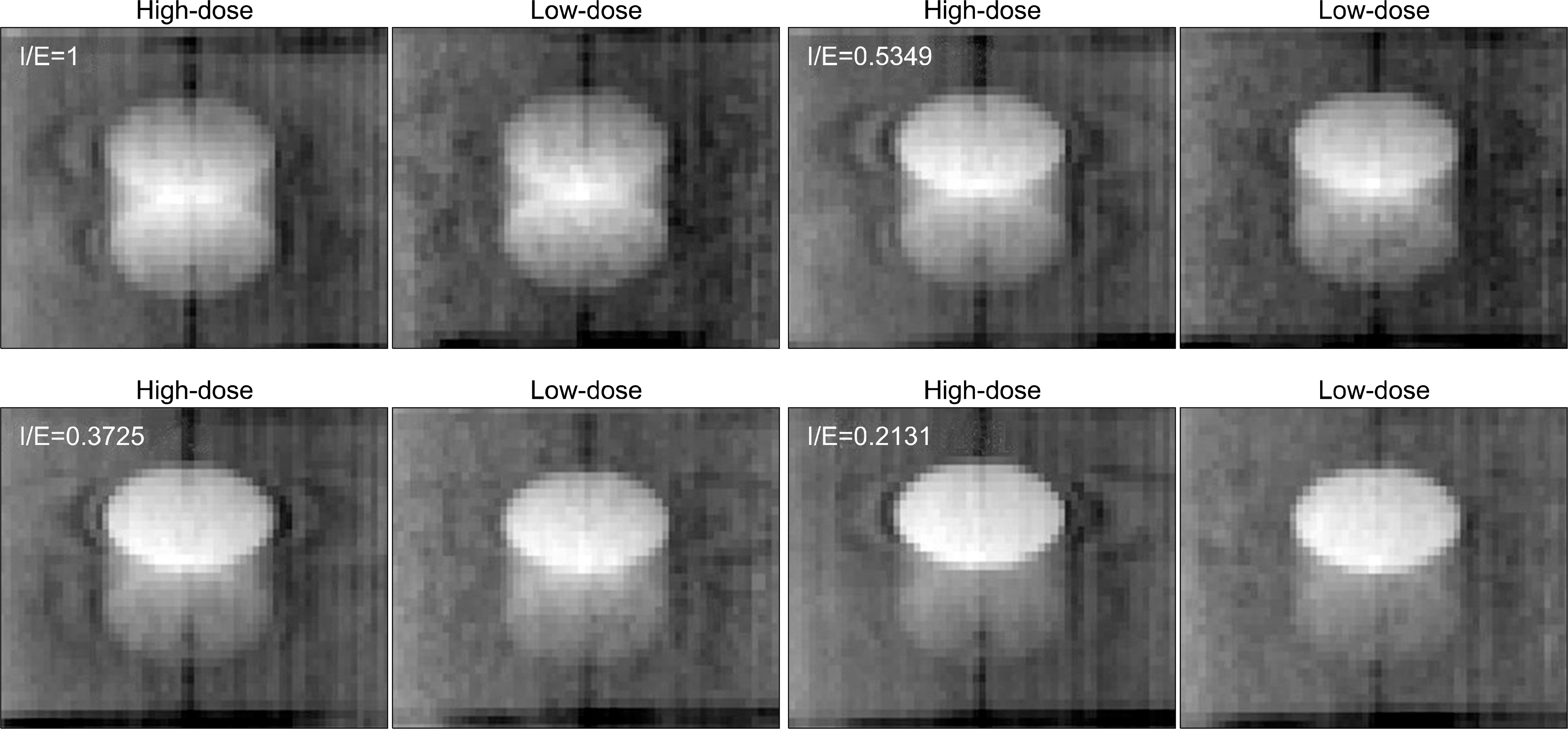
Fig. 5. “I/E Ratio HD_LD Comparisons”. Representative cases of the LIDM and HIDM FBCBCT images illustrating the similarity in ITV visibility between two modes.
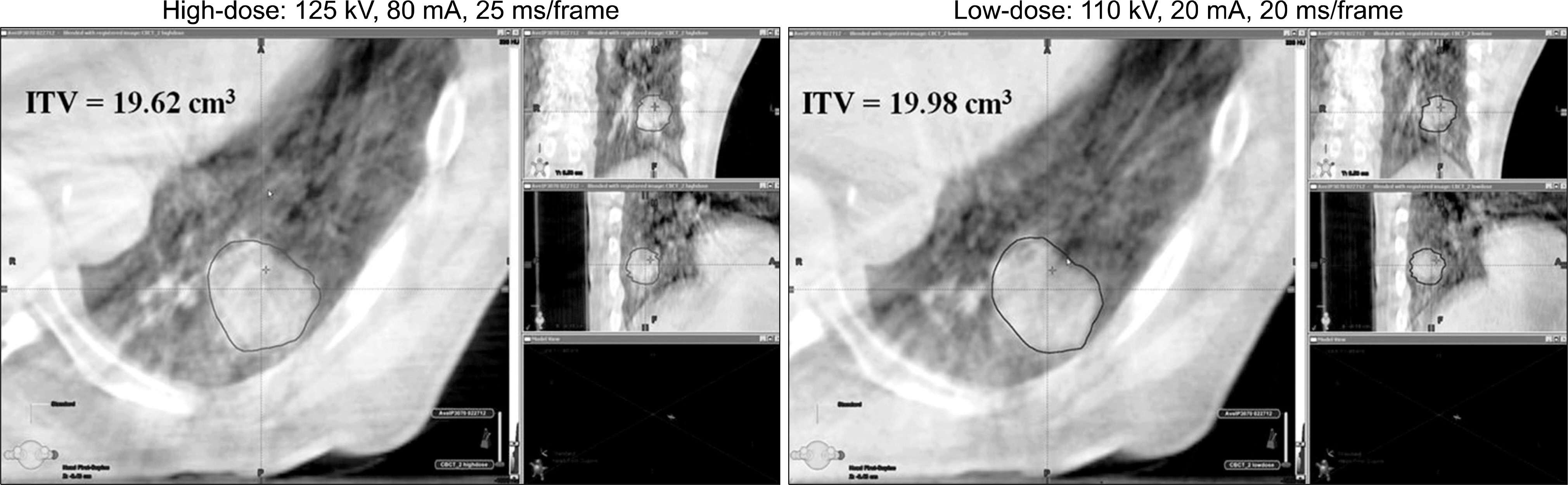
Fig. 6. “HD_LD Contour Comparison”. A representative, example patient case imaged with both the LIDM and HIDM FBCBCTs. An expert Radiation Oncologist contoured the visible ITVs on each image.
Reference
-
1. Vedam SS, Keall PJ, Kini VR, et al. Acquiring a four-dimensional computed tomography dataset using an external respiratory signal. Phys Med Biol. 48(1):45–62. 2003.
Article2. Lu W, Parikh PJ, Hubenschmidt JP, et al. A comparison between amplitude sorting and phase angle sorting using external respiratory measurement for 4DCT. Med Phys. 33(8):2964–2974. 2006.3. Keall PJ. 4-Dimensional computed tomography imaging and treatment planning. Semin Radiat Oncol. 14(1):81–90. 2004.
Article4. Low DA, Nystrom M, Kalinin F, et al. A method for the reconstruction of four-dimensional synchronized CT scans acquired during free breathing. Med Phys. 30(6):1254–1263. 2003.
Article5. Fitzpatrick MJ, Starkschall G, Antolak JA. Displacementbased binning of time-dependent computed tomography image data sets. Med Phys. 33(1):235–246. 2006.
Article6. Wink NM, Panknin C, Soldberg TD. Phase versus amplitude sorting of 4D-CT data. J Appl Clin Med Phys. 7(1):77–85. 2006.
Article7. Ruan D, Fessler JA, Balter JM. Mean position tracking of respiratory motion. Med Phys. 35(2):782–792. 2008.
Article8. Underberg RW, Lagerwaard FJ, Cuijpers JP, et al. Fourdimensional CT scans for treatment planning in stereotactic radiotherapy for stage I lung cancer. Int J Radiat Oncol Biol Phys. 60:1283–1290. 2004.
Article9. Jin JY, Ajlouni M, Chen Q, et al. A technique of using gated- CT images to determine internal target volume (ITV) for fractionated stereotactic lung radiotherapy. Radiother Oncol. 78:177–184. 2006.10. Purdie TG, Bissonnette JP, Franks K, et al. Cone-beam computed tomography for online image guidance of lung stereotactic radiotherapy: Localization, verification, and intrafraction tumor position. Int J Radiat Oncol Biol Phys. 68(1):243–252. 2007.
Article11. Yeung AR, Li JG, Shi W, et al. Tumor localization using cone-beam CT reduces setup margins in conventionally fractionated radiotherapy for lung tumors. Int J Radiat Oncol Biol Phys. 74(4):1100–1107. 2009.
Article12. Grills IS, Hugo G, Kestin LL, et al. Image-guided radiotherapy via daily online cone-beam CT substantially reduces margin requirements for stereotactic lung radiotherapy. Int J Radiat Oncol Biol Phys. 70(4):1045–1056. 2008.
Article13. Yin FF, Wang Z, Yoo S, et al. Integration of cone-beam CT in stereotactic body radiation therapy. Technol Cancer Res Treat. 7(2):133–139. 2008.
Article14. Yin FF, Wang Z, Yoo S, et al. In-room radiographic imaging for localization. Proceedings AAPM Summer School Integrating New Technologies into the Clinic: Monte Carlo and Image Guided Radiation Therapy. 2006, Ontario, Canada.15. Thilmann C, Nill S, Tucking T, et al. Correction of patient positioning errors based on in-line cone beam CTs: clinical implementation and first experiences. Radiother Oncol. 1:16. 2006.
Article16. Li T, Schreibmann E, Yang Y, et al. Motion correction for improved target localization with on-board cone-beam computed tomography. Phys Med Biol. 51:253–267. 2006.
Article17. Duggan DM, Ding GX, Coffey CW, et al. Deep-inspiration breath-hold kilovoltage cone-beam CT for setup of stereotactic body radiation therapy for lung tumors: Initial experience. Lung Cancer. 56(1):77–88. 2007.
Article18. Yin FF, Das S, Kirkpatrick J, et al. Physics and imaging for targeting of oligometastases. Semin Radiat Oncol. 16(2):85–101. 2006.
Article19. Vergalasova I, Maurer J, Yin FF. Potential underestimation of the internal target volume (ITV) from free-breathing CBCT. Med Phys. 38(8):4689–4699. 2011.
Article20. Buzug TM. Computed Tomography: From Photon Statistics to Modern Cone-Beam CT. 1st ed. Springer-Verlag Berlin, Heidelberg;2008. p. 521.21. Hsieh J. Computed Tomography: Principles, Design, Artifacts, and Recent Advances. 2nd ed. SPIE/John Wiley & Songs, Inc.;2009. p. 556.22. Li T, Xing L. Optimizing 4D cone-beam CT acquisition protocol for external beam radiotherapy. Int J Radiat Oncol Biol Phys. 67(4):1211–1219. 2007.
Article23. Song WY, Kamath S, Ozawa S, et al. A dose comparison study between XVI and OBI CBCT systems. Med Phys. 35(2):480–486. 2008.24. Sonke JJ, Zijp L, Remeijer P, et al. Respiratory correlated cone beam CT. Med Phys. 32(4):1176–1186. 2005.
Article25. Nakayama Y, Awai K, Funama Y, et al. Abdominal CT with low Tube Voltage: Preliminary Observations about Radiation Dose, Contrast Enhancement, Image Quality, and Noise. Radiology. 237(3):945–951. 2005.
Article26. Hsieh J. Adaptive streak artifact reduction in computed tomography resulting from excessive x-ray photon noise. Med Phys. 25(11):2139–2147. 1998.
Article
- Full Text Links
-
- Actions
-
Cited
- CITED
-
- Close
- Share
-
- Similar articles
-
- Practical Considerations in Preparing an Institutional Procedure of Image Guided Radiation Therapy
- Analysis of the priority of anatomic structures according to the diagnostic task in cone-beam computed tomographic images
- Efficacy of a Respiratory Training System on the Regularity of Breathing
- On-line Image Guided Radiation Therapy using Cone-Beam CT (CBCT)
- Half-Fan-Based Intensity-Weighted Region-of-Interest Imaging for Low-Dose Cone-Beam CT in Image-Guided Radiation Therapy
