Half-Fan-Based Intensity-Weighted Region-of-Interest Imaging for Low-Dose Cone-Beam CT in Image-Guided Radiation Therapy
- Affiliations
-
- 1Department of Nuclear and Quantum Engineering, Korea Advanced Institute of Science and Technology, Daejeon, Korea. scho@kaist.ac.kr
- 2Department of Radiation Oncology, Yonsei Cancer Center, Yonsei University College of Medicine, Seoul, Korea.
- 3Department of Nuclear Engineering, Khalifa University, Abu Dhabi, UAE.
- KMID: 2357386
- DOI: http://doi.org/10.4258/hir.2016.22.4.316
Abstract
OBJECTIVES
With the increased use of computed tomography (CT) in clinics, dose reduction is the most important feature people seek when considering new CT techniques or applications. We developed an intensity-weighted region-of-interest (IWROI) imaging method in an exact half-fan geometry to reduce the imaging radiation dose to patients in cone-beam CT (CBCT) for image-guided radiation therapy (IGRT). While dose reduction is highly desirable, preserving the high-quality images of the ROI is also important for target localization in IGRT.
METHODS
An intensity-weighting (IW) filter made of copper was mounted in place of a bowtie filter on the X-ray tube unit of an on-board imager (OBI) system such that the filter can substantially reduce radiation exposure to the outer ROI. In addition to mounting the IW filter, the lead-blade collimation of the OBI was adjusted to produce an exact half-fan scanning geometry for a further reduction of the radiation dose. The chord-based rebinned backprojection-filtration (BPF) algorithm in circular CBCT was implemented for image reconstruction, and a humanoid pelvis phantom was used for the IWROI imaging experiment.
RESULTS
The IWROI image of the phantom was successfully reconstructed after beam-quality correction, and it was registered to the reference image within an acceptable level of tolerance. Dosimetric measurements revealed that the dose is reduced by approximately 61% in the inner ROI and by 73% in the outer ROI compared to the conventional bowtie filter-based half-fan scan.
CONCLUSIONS
The IWROI method substantially reduces the imaging radiation dose and provides reconstructed images with an acceptable level of quality for patient setup and target localization. The proposed half-fan-based IWROI imaging technique can add a valuable option to CBCT in IGRT applications.
Keyword
MeSH Terms
Figure
-

Figure 1 Intensity-weighted region-of-interest (IWROI) imaging schematic in a conventional half-fan geometry. FOV: field-of-view.

Figure 2 Schematic of a rebinned half-fan geometry.
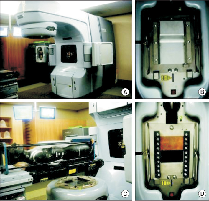
Figure 3 Images of (A) an on-board imager (OBI) system, (B) a mounted half-fan bowtie filter, (C) a humanoid phantom, and (D) a mounted intensity-weighting filter.
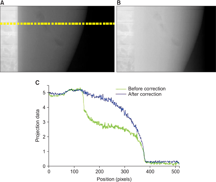
Figure 4 Log-transformed projection data (A) before and (B) after beam-quality correction. (C) Line profiles are shown for comparison. Note that the off-axis data are not shown.

Figure 5 (A) An open-field intensity map and (B) a raw projection of a phantom.

Figure 6 Film and glass dosimeters placed in the phantom for dosimetric measurements. XRCT: X-ray computed tomography.
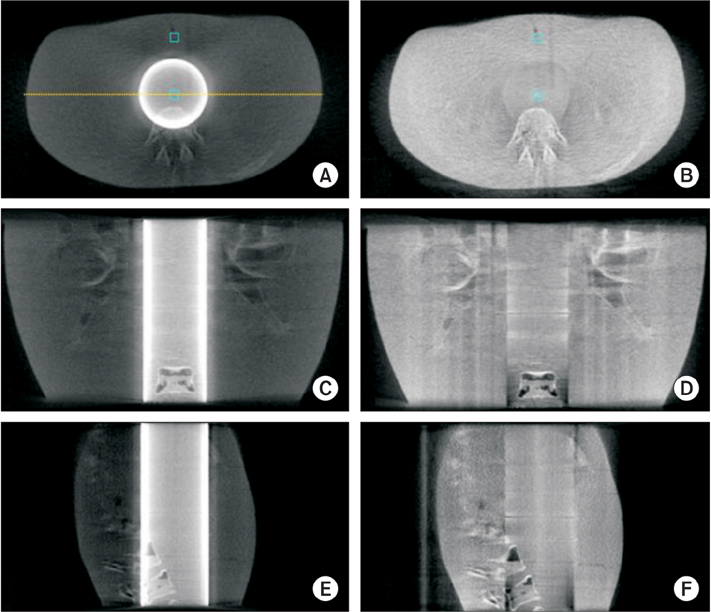
Figure 7 Transverse images (A) before and (B) after; coronal images (C) before and (D) after; and sagittal images (E) before and (F) after beam-quality correction. The display window is [10, 250] HU for the images on the left and [10, 160] HU for the images on the right.

Figure 8 Line profiles of two images along the dashed line shown in Figure 7A. The solid line represents the uncorrected image and dashed line represents the corrected case.

Figure 9 Transverse slice of the target and moving images on a checkerboard: (A) before and (B) after image registration.
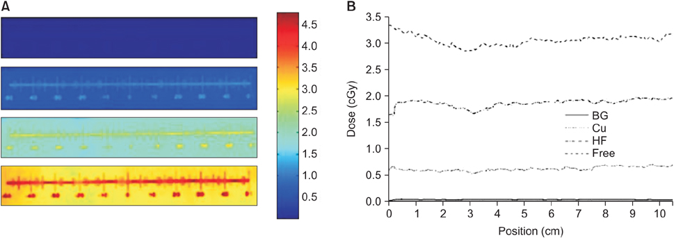
Figure 10 (A) Two-dimensional distribution of the imaging radiation dose (cGy) within the XRCT film for various experiments: no radiation or background (top), proposed method using a Cu filter (middle high), half-fan bowtie filter (middle low), and filter-free case (bottom). (B) Corresponding line profiles along the horizontal line near the bottom edge of the XRCT films are also shown. XRCT: X-ray computed tomography.
Reference
-
1. Chen GT, Sharp GC, Mori S. A review of image-guided radiotherapy. Radiol Phys Technol. 2009; 2(1):1–12.
Article2. Murphy MJ, Balter J, Balter S, BenComo JA Jr, Das IJ, Jiang SB, et al. The management of imaging dose during image-guided radiotherapy: report of the AAPM Task Group 75. Med Phys. 2007; 34(10):4041–4063.
Article3. Sidky EY, Kao CM, Pan X. Accurate image reconstruction from few-views and limited-angle data in divergent-beam CT. J Xray Sci Technol. 2006; 14(2):119–139.4. Sidky EY, Pan X. Image reconstruction in circular cone-beam computed tomography by constrained, total-variation minimization. Phys Med Biol. 2008; 53(17):4777–4807.
Article5. Bian J, Siewerdsen JH, Han X, Sidky EY, Prince JL, Pelizzari CA, et al. Evaluation of sparse-view reconstruction from flat-panel-detector cone-beam CT. Phys Med Biol. 2010; 55(22):6575–6599.
Article6. Wang J, Li T, Xing L. Iterative image reconstruction for CBCT using edge-preserving prior. Med Phys. 2009; 36(1):252–260.
Article7. Jia X, Lou Y, Li R, Song WY, Jiang SB. GPU-based fast cone beam CT reconstruction from undersampled and noisy projection data via total variation. Med Phys. 2010; 37(4):1757–1760.
Article8. Yan H, Cervino L, Jia X, Jiang SB. A comprehensive study on the relationship between the image quality and imaging dose in low-dose cone beam CT. Phys Med Biol. 2012; 57(7):2063–2080.
Article9. Chityala RN, Hoffmann KR, Bednarek DR, Rudin S. Region of interest (ROI) computed tomography. Proc SPIE Int Soc Opt Eng. 2004; 5368:534–541.
Article10. Chen L, Shaw CC, Altunbas MC, Lai CJ, Liu X, Han T, et al. Feasibility of volume-of-interest (VOI) scanning technique in cone beam breast CT: a preliminary study. Med Phys. 2008; 35(8):3482–3490.
Article11. Cho S, Pearson E, Pelizzari CA, Pan X. Region-of-interest image reconstruction with intensity weighting in circular cone-beam CT for image-guided radiation therapy. Med Phys. 2009; 36(4):1184–1192.
Article12. Schafer S, Noel PB, Walczak AM, Hoffmann KR. Filtered region of interest cone-beam rotational angiography. Med Phys. 2010; 37(2):694–703.
Article13. Kolditz D, Kyriakou Y, Kalender WA. Volume-of-interest (VOI) imaging in C-arm flat-detector CT for high image quality at reduced dose. Med Phys. 2010; 37(6):2719–2730.
Article14. Zou Y, Pan X, Sidky EY. Image reconstruction in regions-of-interest from truncated projections in a reduced fan-beam scan. Phys Med Biol. 2005; 50(1):13–27.
Article15. Cho S, Bian J, Pelizzari CA, Chen CT, He TC, Pan X. Region-of-interest image reconstruction in circular cone-beam microCT. Med Phys. 2007; 34(12):4923–4933.
Article16. Cho PS, Rudd AD, Johnson RH. Cone-beam CT from width-truncated projections. Comput Med Imaging Graph. 1996; 20(1):49–57.
Article17. Ohnesorge B, Flohr T, Schwarz K, Heiken JP, Bae KT. Efficient correction for CT image artifacts caused by objects extending outside the scan field of view. Med Phys. 2000; 27(1):39–46.
Article18. Feldkamp LA, Davis LC, Kress JW. Practical cone-beam algorithm. J Opt Soc Am A. 1984; 1(6):612–619.
Article19. Yu L, Pelizzari C, Pan X, Riem H, Munro P, Kaissl W. Application of asymmetric cone-beam CT in radiotherapy. In : Proceedings of 2004 IEEE Nuclear Science Symposium Conference Record; 2004 Oct 16-22; Rome. Italy: p. 3249–3252.20. Leng S, Zhuang T, Nett BE, Chen GH. Exact fan-beam image reconstruction algorithm for truncated projection data acquired from an asymmetric half-size detector. Phys Med Biol. 2005; 50(8):1805–1820.
Article21. Zou Y, Pan X. Image reconstruction on PI-lines by use of filtered backprojection in helical cone-beam CT. Phys Med Biol. 2004; 49(12):2717–2731.
Article22. Li L, Chen Z, Zhang L, Xing Y, Kang K. A cone-beam tomography system with a reduced size planar detector: a backprojection-filtration reconstruction algorithm as well as numerical and practical experiments. Appl Radiat Isot. 2007; 65(9):1041–1047.
Article23. Yu L, Xia D, Zou Y, Sidky EY, Bian J, Pan X. A rebinned backprojection-filtration algorithm for image reconstruction in helical cone-beam CT. Phys Med Biol. 2007; 52(18):5497–5508.
Article24. Turbell H. Cone-beam reconstruction using filtered backprojection [dissertation]. Linkoping, Sweden: Linkoping University;2001.25. Schafer D, Grass M, van de Haar P. FBP and BPF reconstruction methods for circular X-ray tomography with off-center detector. Med Phys. 2011; 38:Suppl 1. S85.26. Wang J, Li T, Lu H, Liang Z. Penalized weighted least-squares approach to sinogram noise reduction and image reconstruction for low-dose X-ray computed tomography. IEEE Trans Med Imaging. 2006; 25(10):1272–1283.
Article27. Wang J, Zhu L, Xing L. Noise reduction in low-dose x-ray fluoroscopy for image-guided radiation therapy. Int J Radiat Oncol Biol Phys. 2009; 74(2):637–643.
Article
- Full Text Links
-
- Actions
-
Cited
- CITED
-
- Close
- Share
-
- Similar articles
-
- Practical Considerations in Preparing an Institutional Procedure of Image Guided Radiation Therapy
- On-line Image Guided Radiation Therapy using Cone-Beam CT (CBCT)
- Improvement of the Dose Calculation Accuracy Using MVCBCT Image Processing
- Error Analysis of Delivered Dose Reconstruction Using Cone-beam CT and MLC Log Data
- On-line Setup for Lung Cancer Patient in Stereotactic Radiation Surgery using CBCT
