Eight prehilar branches of the right renal artery
- Affiliations
-
- 1Department of Anatomy, Melaka Manipal Medical College (Manipal Campus), Manipal University, Manipal, Karnataka, India. nayaksathish@gmail.com
- KMID: 1882599
- DOI: http://doi.org/10.5115/acb.2014.47.3.214
Abstract
- Imaging technology with its advancement in the field of urology is the boon for the patients who require minimally invasive approaches for various kidney disorders. These approaches require a precise knowledge of the normal and variant anatomy of vessels at the hilum of the kidney. During routine dissections, a variation in the branching pattern of the right renal artery was noted in an adult male cadaver. The right renal artery divided into upper and lower divisions 6cm away from the hilum of the kidney. The upper division gave 4 branches, and the lower division gave two branches. These two branches further bifurcated and gave 2 branches each. Thus, there were 8 prehilar branches of renal artery. The multiple prehilar branches led to a congested atmosphere at the hilum of the kidney. This arterial congestion might result in hindering the blood flow at the renal hilum. Apart from this, it might cause difficulties in diagnostic and therapeutic invasive procedures. Knowledge of this variation is of importance to radiologists and urologists in particular.
Keyword
MeSH Terms
Figure
-

Fig. 1 Anterior view of the renal hilum showing the branches of right renal artery. AA, aberrant branches; IVC, inferior vena cava; LD, lower division of renal artery; RK, right kidney; RISA, right inferior suprarenal artery; RRA, right renal artery; RSG, right suprarenal gland; UD, upper division of the renal artery.

Fig. 2 Anterior view of the renal hilum showing all the eight branches of right renal artery. AA, abdominal aorta; IVC, inferior vena cava; LD, lower division of renal artery; RK, right kidney; RRA, right renal artery; RRV, right renal vein; RSG, right suprarenal gland; UD, upper division of the renal artery.
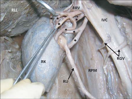
Fig. 3 Anterior inferior view of the renal hilum showing the branches of renal artery (asterisks). IVC, inferior vena cava; RGV, right gonadal vessels; RLL, right lobe of liver; RK, right kidney; RPM, right psoas major; RRV, right renal vein; RU, right ureter.
Reference
-
1. Standring S. Gray's anatomy: the anatomical basis of clinical practice. 39th ed. New York: Churchill Livingstone, Elsevier;2005. p. 1271–1274.2. Satyapal KS, Haffejee AA, Singh B, Ramsaroop L, Robbs JV, Kalideen JM. Additional renal arteries: incidence and morphometry. Surg Radiol Anat. 2001; 23:33–38.3. Bayramoglu A, Demiryurek D, Erbil KM. Bilateral additional renal arteries and an additional right renal vein associated with unrotated kidneys. Saudi Med J. 2003; 24:535–537.4. Ozkan U, Oğuzkurt L, Tercan F, Kizilkiliç O, Koç Z, Koca N. Renal artery origins and variations: angiographic evaluation of 855 consecutive patients. Diagn Interv Radiol. 2006; 12:183–186.5. Nayak BS. Multiple variations of the right renal vessels. Singapore Med J. 2008; 49:e153–e155.6. Kumar N, Aithal AP, Guru A, Nayak SB. Bilateral vascular variations at the renal hilum: a case report. Case Rep Vasc Med. 2012; 2012:968506.7. Rouvière O, Lyonnet D, Berger P, Pangaud C, Gelet A, Martin X. Ureteropelvic junction obstruction: use of helical CT for preoperative assessment: comparison with intraarterial angiography. Radiology. 1999; 213:668–673.8. Das S, Paul S. Variation of renal hilar structures: a cadaveric case. Eur J Anat. 2006; 10:41–43.9. Rapp DE, Orvieto MA, Gerber GS, Johnston WK 3rd, Wolf JS Jr, Shalhav AL. En bloc stapling of renal hilum during laparoscopic nephrectomy and nephroureterectomy. Urology. 2004; 64:655–659.
- Full Text Links
-
- Actions
-
Cited
- CITED
-
- Close
- Share
-
- Similar articles
-
- Establishment of normal reference of radiological morphology of renal artery in mini-pigs by renal angiography
- Accessory Renal Arteries Found during Dissection
- A Case of Curable Hypertension Owing to Unilateral Renal Hypoplasia
- Hybrid Treatment of Coexisting Renal Artery Aneurysm and Abdominal Aortic Aneurysm in a Gallbladder Cancer Patient
- Normal variations of renal vessels based upon the study of 240 living-donor nephrectomies
