Anat Cell Biol.
2014 Sep;47(3):202-206. 10.5115/acb.2014.47.3.202.
Mastoid emissary foramina: an anatomical morphological study with discussion on their evolutionary and clinical implications
- Affiliations
-
- 1Department of Anatomy, Kasturba Medical College, Manipal University, Mangalore, India. flutesnowmm@gmail.com
- 2Department of Anatomy, Melaka Manipal Medical College, Manipal University, Manipal, India.
- KMID: 1882596
- DOI: http://doi.org/10.5115/acb.2014.47.3.202
Abstract
- The identification of mastoidal emissary veins is of importance in the neurosurgical practice to diagnose abnormal and normal structures. In the present study, the objectives were to estimate the prevalence rate of mastoidal emissary foramina in the temporal bones of the adult skull and to study their number and morphology. The present study included 48 adult human skulls which were obtained from the gross anatomy laboratory of our institution. The mastoid parts of 96 temporal bones were macroscopically observed for the prevalence, number and morphology of the emissary foramina. It is observed that, the mastoidal emissary foramen was present in 88 temporal bones (91.7%) of our specimens. The foramen was observed single in 60 temporal bones (62.5%), double in 22 bones (22.9%), and triple in 6 temporal bones (6.2%). The mastoidal emissary foramen was absent in 8 (8.3%) temporal bones. The foramen was bilaterally absent in 3 (3.1%) skulls. It was unilaterally absent in 2 (2.1%) skulls and both were on the left side. The mastoidal emissary vein is prevalent in a large number (91.7%) of cases. It was observed that the accessory mastoidal emissary foramina were present in 29.1% of cases. Recognition of the mastoid emissary veins and accessory mastoid emissary veins during the otologic surgery is critical to avoid the significant bleeding. In the neurosurgical practice, the knowledge is important due to variability in the number of mastoidal emissary veins and their connection to the venous sinuses.
Keyword
Figure
-

Fig. 1 Morphological distribution of the mastoid emissary foramina observed in the present study (n=96).
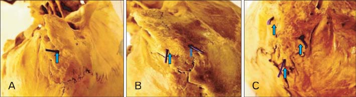
Fig. 2 Pictures of the skull bases of the present study showing. (A) Single mastoidal emissary foramen (62.5%). (B) Double mastoidal emissary foramen (22.9%). (C) Triple mastoidal emissary foramen (6.2%).
Cited by 1 articles
-
Incidental occurrence of an unusually large mastoid foramen on cone-beam computed tomography and review of the literature
Ali Z. Syed, Cleo Sin, Raquel Rios, Mel Mupparapu
Imaging Sci Dent. 2016;46(1):39-45. doi: 10.5624/isd.2016.46.1.39.
Reference
-
1. Singhal S, Ravindranath R. Occipital emissary foramina in South Indian modern human skulls. ISRN Anat. 2013; 2013:727489.2. Sicher H, DuBrul EL. Oral anatomy. 6th ed. Rio de Janeiro: Guanabara Koogan;1977.3. Shaik HS, Shepur MP, Desai SD, Thomas ST, Maavishettar GF, Haseena S. Study of mastoid canals and grooves in South Indian skulls. Indian J Med Healthc. 2012; 1:32–33.4. Louis RG Jr, Loukas M, Wartmann CT, Tubbs RS, Apaydin N, Gupta AA, Spentzouris G, Ysique JR. Clinical anatomy of the mastoid and occipital emissary veins in a large series. Surg Radiol Anat. 2009; 31:139–144.5. El Kettani C, Badaoui R, Fikri M, Jeanjean P, Montpellier D, Tchaoussoff J. Pulmonary oedema after venous air embolism during craniotomy. Eur J Anaesthesiol. 2002; 19:846–848.6. Reis CV, Deshmukh V, Zabramski JM, Crusius M, Desmukh P, Spetzler RF, Preul MC. Anatomy of the mastoid emissary vein and venous system of the posterior neck region: neurosurgical implications. Neurosurgery. 2007; 61:5 Suppl 2. 193–200.7. Pereira GA, Lopes PT, Santos AM, Pozzobon A. Study of landmarks in dried skulls in a Brazil population. J Morphol Sci. 2013; 30:94–97.8. Singh R. Morphometric analysis of infraorbital foramen in Indian dry skulls. Anat Cell Biol. 2011; 44:79–83.9. Kim LK, Ahn CS, Fernandes AE. Mastoid emissary vein: anatomy and clinical relevance in plastic & reconstructive surgery. J Plast Reconstr Aesthet Surg. 2014; 67:775–780.10. Falk D, Conroy GC. The cranial venous sinus system in Australopithecus afarensis. Nature. 1983; 306:779–781.11. Falk D. Evolution of cranial blood drainage in hominids: enlarged occipital/marginal sinuses and emissary foramina. Am J Phys Anthropol. 1986; 70:311–324.12. Kimbel WH. Variation in the pattern of cranial venous sinuses and hominid phylogeny. Am J Phys Anthropol. 1984; 63:243–263.13. Goucha S, Mnif N, Bouhala T, Tenzakhti F, El Andaloussi H, Fazaa B, Hamza R, Kamoun MR. Value of imaging in GAPO syndrome. J Radiol. 2002; 83(2 Pt 1):153–156.14. Mavrodi A, Paraskevas G. Evolution of the paranasal sinuses' anatomy through the ages. Anat Cell Biol. 2013; 46:235–238.15. Kaur J, Srivastava D, Singh D, Raheja S. The study of hyperostosic variants: significance of hyperostotic variants of human skulls in anthropology. Anat Cell Biol. 2012; 45:268–273.16. Keskil S, Gözil R, Calgüner E. Common surgical pitfalls in the skull. Surg Neurol. 2003; 59:228–231.17. Freire AR, Rossi AC, de Oliveira VC, Prado FB, Caria PH, Botacin PR. Emissary foramens of the human skull: anatomical characteristics and its relations with clinical neurosurgery. Int J Morphol. 2013; 31:287–292.18. Gözil R, Kadioglu D, Calgüner E. Occipital emissary foramen in skulls from central Anatolia. Acta Anat (Basel). 1995; 153:325–326.19. Berge JK, Bergman RA. Variations in size and in symmetry of foramina of the human skull. Clin Anat. 2001; 14:406–413.20. Wysocki J, Reymond J, Skarzyński H, Wróbel B. The size of selected human skull foramina in relation to skull capacity. Folia Morphol (Warsz). 2006; 65:301–308.21. Boyd GI. The emissary foramina of the cranium in man and the anthropoids. J Anat. 1930; 65(Pt 1):108–121.22. Friedmann DR, Amoils M, Germiller JA, Lustig LR, Glastonbury CM, Pramanik BK, Lalwani AK. Venous malformations of the temporal bone are a common feature in CHARGE syndrome. Laryngoscope. 2012; 122:895–900.23. Cheatle A. The mastoid emissary vein and its surgical importance. Proc R Soc Med. 1925; 18:29–34.24. Murlimanju BV, Prabhu LV, Pai MM, Jaffar M, Saralaya VV, Tonse M, M D P. Occipital emissary foramina in human skulls: an anatomical investigation with reference to surgical anatomy of emissary veins. Turk Neurosurg. 2011; 21:36–38.
- Full Text Links
-
- Actions
-
Cited
- CITED
-
- Close
- Share
-
- Similar articles
-
- Morphology and topography of the parietal emissary foramina in South Indians: an anatomical study
- Pulsatile Tinnitus Caused by a Dilated Mastoid Emissary Vein
- Mastoid Foramen and Superficial Mastoid Canals of Korean Men
- Non-Metrical Morphologic Variations of Korean Skull Foramina
- Anatomical structure of lingual foramen in cone beam computed tomography
