Pulsatile Tinnitus Caused by a Dilated Mastoid Emissary Vein
- Affiliations
-
- 1Department of Neurology, School of Medicine, Kangwon National University, Chuncheon, Korea.
- 2Department of Radiology, School of Medicine, Kangwon National University, Chuncheon, Korea.
- 3Department of Plastic and Reconstructive Surgery, School of Medicine, Kangwon National University, Chuncheon, Korea.
- 4Department of Otolaryngology, School of Medicine, Kangwon National University, Chuncheon, Korea. birdynec@kangwon.ac.kr
- KMID: 1786975
- DOI: http://doi.org/10.3346/jkms.2013.28.4.628
Abstract
- Although pulsatile tinnitus can be audible, objective demonstration of this heartbeat-synchronous sound has rarely been successful. We report a rare case of pulsatile tinnitus in a 44-yr-old female patient, which was induced by a large mastoid emissary vein (MEV) and objectively documented by Doppler sonography of the left posterior auricular region. The tinnitus was intermittent and the patient could adapt to the tinnitus without intervention on the mastoid emissary vein. These findings suggest that a single large MEV can cause pulsatile tinnitus in the absence of other vascular abnormalities, and imaging studies of the posterior fossa and Doppler ultrasonography can aid the diagnosis in such cases.
Keyword
MeSH Terms
Figure
-
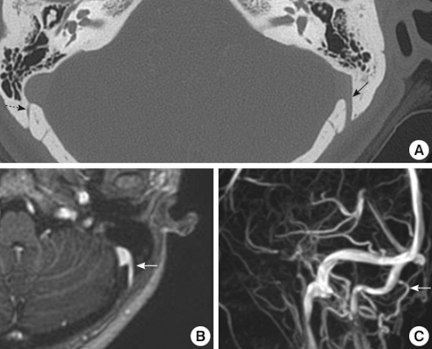
Fig. 1 Imaging studies revealing the large mastoid emissary vein (MEV). The patient's temporal bone CT scan showed a prominent orifice of the large left MEV (black arrow), with a diameter of 4.5 mm at the inner foramen, abnormally larger than the other side (dotted arrow) (A). The enhanced MRI and MR venogram showed blood flow of the left MEV draining into the sigmoid sinus (white arrows) (B, C).
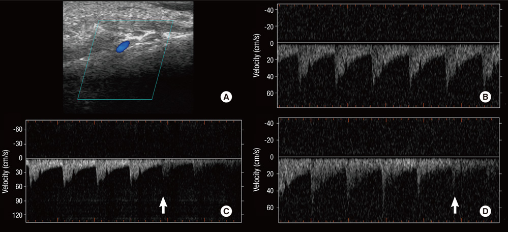
Fig. 2 Doppler sonogram of the pulsating flow in the mastoid emissary vein (MEV), correlating to the pulsatile tinnitus. Doppler sonography identified the location of the outer foramen of the MEV and the pulsating venous blood flow draining into the sigmoid sinus (A, B). The patient recognized reduction of the pulsatile tinnitus synchronously with the decrease of the flow-signal by compression of the left jugular vein (C) and the Valsalva maneuver (D). Arrows: starting points of compression of the left jugular vein and the Valsalva maneuver, respectively.
Cited by 1 articles
-
Incidental occurrence of an unusually large mastoid foramen on cone-beam computed tomography and review of the literature
Ali Z. Syed, Cleo Sin, Raquel Rios, Mel Mupparapu
Imaging Sci Dent. 2016;46(1):39-45. doi: 10.5624/isd.2016.46.1.39.
Reference
-
1. Lo WW, Solti-Bohman LG, McElveen JT Jr. Aberrant carotid artery: radiologic diagnosis with emphasis on high-resolution computed tomography. Radiographics. 1985. 5:985–993.2. Levine SB, Snow JB Jr. Pulsatile tinnitus. Laryngoscope. 1987. 97:401–406.3. Dietz RR, Davis WL, Harnsberger HR, Jacobs JM, Blatter DD. MR imaging and MR angiography in the evaluation of pulsatile tinnitus. AJNR Am J Neuroradiol. 1994. 15:879–889.4. Forte V, Turner A, Liu P. Objective tinnitus associated with abnormal mastoid emissary vein. J Otolaryngol. 1989. 18:232–235.5. Louis RG Jr, Loukas M, Wartmann CT, Tubbs RS, Apaydin N, Gupta AA, Spentzouris G, Ysique JR. Clinical anatomy of the mastoid and occipital emissary veins in a large series. Surg Radiol Anat. 2009. 31:139–144.6. San Millán Ruíz D, Gailloud P, Rüfenacht DA, Delavelle J, Henry F, Fasel JH. The craniocervical venous system in relation to cerebral venous drainage. AJNR Am J Neuroradiol. 2002. 23:1500–1508.7. Hoffmann O, Klingebiel R, Braun JS, Katchanov J, Valdueza JM. Diagnostic pitfall: atypical cerebral venous drainage via the vertebral venous system. AJNR Am J Neuroradiol. 2002. 23:408–411.8. Kudo K, Terae S, Ishii A, Omatsu T, Asano T, Tha KK, Miyasaka K. Physiologic change in flow velocity and direction of dural venous sinuses with respiration: MR venography and flow analysis. AJNR Am J Neuroradiol. 2004. 25:551–557.9. Chauhan NS, Sharma YP, Bhagra T, Sud B. Persistence of multiple emissary veins of posterior fossa with unusual origin of left petrosquamosal sinus from mastoid emissary. Surg Radiol Anat. 2011. 33:827–831.
- Full Text Links
-
- Actions
-
Cited
- CITED
-
- Close
- Share
-
- Similar articles
-
- A Case of Pulsatile Tinnitus from Dehiscent High Jugular Bulb Treated by Reconstruction of the Hypotympanum
- Mastoid emissary foramina: an anatomical morphological study with discussion on their evolutionary and clinical implications
- A Case of Pulsatile Tinnitus with High Jugular Bulb Treated by Ligation of Internal Jugular Vein
- Pulsatile Tinnitus Caused by Arteriovenous Fistula of External Carotid Artery
- A Case of Jugular Bulb Diverticulum Accompanied with Pulsatile Tinnitus
