Anat Cell Biol.
2015 Dec;48(4):292-298. 10.5115/acb.2015.48.4.292.
Morphology and topography of the parietal emissary foramina in South Indians: an anatomical study
- Affiliations
-
- 1Department of Anatomy, Kasturba Medical College, Manipal University, Mangalore, Karnataka, India. flutemist@gmail.com
- 2Department of Anatomy, Srinivas Institute of Medical Sciences & Research Centre, Srinivas University, Mangalore, Karnataka, India.
- KMID: 2133270
- DOI: http://doi.org/10.5115/acb.2015.48.4.292
Abstract
- The objectives of the present study were to study the prevalence of the parietal emissary vein in adult South Indian population and to study the distance of foramen from the sagittal suture. There were 58 adult human skulls in the present study which were available at the anatomy department of our institution. The study included 116 parietal bones which have been observed macroscopically for the number, prevalence and topography of the emissary foramen. The emissary foramen was present in 83 parietal bones (71.5%) of the present study. It was present at the junction between the middle 1/3 and posterior 1/3 region of the parietal bone. The foramen was observed solitary in 73 parietal bones (62.9%), double in 8 bones (6.9%), and triple in 2 parietal bones (1.7%). The foramen was not observed in 33 parietal bones (28.4%). The bilateral absence of parietal emissary foramen was seen in 7 skulls (12.1%). It was absent unilaterally in 19 skulls (32.7%). The accessory foramina were seen in only 8 skulls (13.8%). The mean distance of the foramen from the sagittal suture was 6.7+/-2.9 mm and 6.8+/-2.8 mm on the right and left sides respectively. The prevalence of parietal emissary vein in the present study was 71.5%. The present study has observed important data about the morphology and morphometry of the parietal emissary vein in South Indian population. The identification of parietal emissary veins and accessory veins is important in the operation room to prevent the blood loss.
Keyword
Figure
-
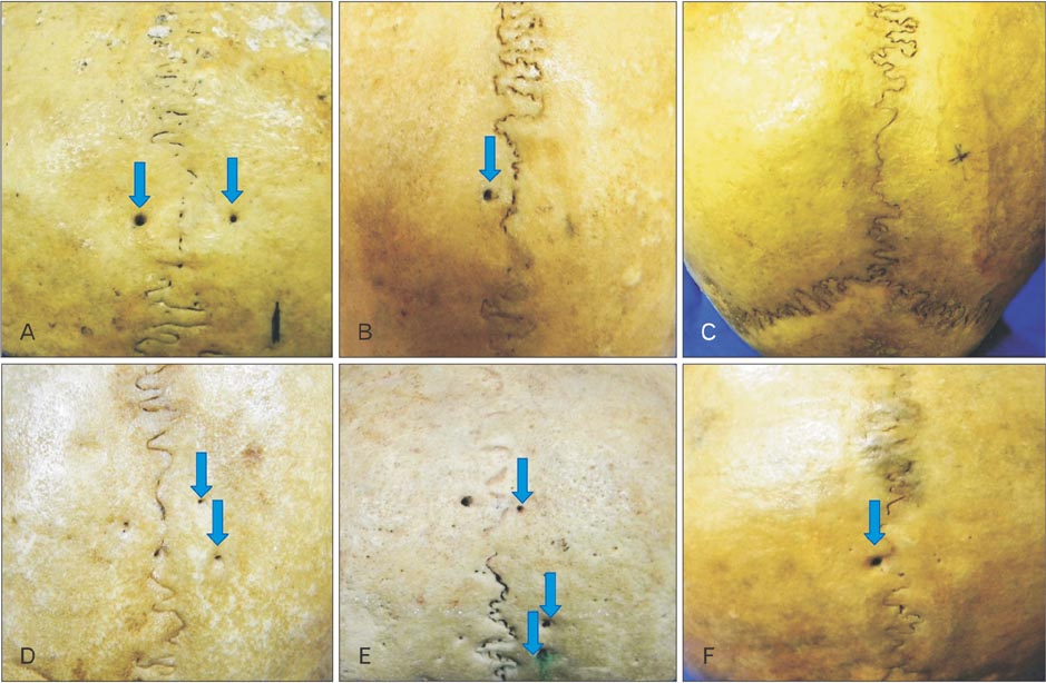
Fig. 1 The crania of the present study. (A) Bilateral presence (arrows) of the parietal emissary foramen (55.2%). (B) The unilateral presence (arrow) of the parietal emissary foramen (32.7%). (C) The bilateral absence of the parietal emissary foramen (12.1%). (D) Right parietal bone showing the double (arrows) emissary foramina (3.5%). (E) Right parietal bone showing the triple (arrows) emissary foramina (0.9%). (F) The parietal emissary foramen (arrow) over the sagittal suture (3.4%).
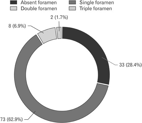
Fig. 2 The frequency of distribution of the number of emissary foramen in the parietal bones of the present study (n=116).
Reference
-
1. Currarino G. Normal variants and congenital anomalies in the region of the obelion. AJR Am J Roentgenol. 1976; 127:487–494.2. Freire AR, Rossi AC, De Oliveira VC, Prado FB, Caria PH, Botacin PR. Emissary foramens of the human skull: anatomical characteristics and its relations with clinical neurosurgery. Int J Morphol. 2013; 31:287–292.3. Jamieson EB. Dixon's manual of human osteology. 2nd ed. London: Humphrey Milford;1937.4. Wysocki J, Reymond J, Skarzyński H, Wróobel B. The size of selected human skull foramina in relation to skull capacity. Folia Morphol (Warsz). 2006; 65:301–308.5. Yoshioka N, Rhoton AL Jr, Abe H. Scalp to meningeal arterial anastomosis in the parietal foramen. Neurosurgery. 2006; 58:ONS123–ONS126.6. Boyd GI. The emissary foramina of the cranium in man and the anthropoids. J Anat. 1930; 65:108–121.7. Murlimanju BV, Chettiar GK, Prameela MD, Tonse M, Kumar N, Saralaya VV, Prabhu LV. Mastoid emissary foramina: an anatomical morphological study with discussion on their evolutionary and clinical implications. Anat Cell Biol. 2014; 47:202–206.8. Murlimanju BV, Prabhu LV, Pai MM, Jaffar M, Saralaya VV, Tonse M, M DP. Occipital emissary foramina in human skulls: an anatomical investigation with reference to surgical anatomy of emissary veins. Turk Neurosurg. 2011; 21:36–38.9. Murlimanju BV, Reddy GR, Latha VP, Vasudha VS, Rao CP, Mangala MP, Ashwin K, Rajanigandha V. Foramen of Vesalius: prevalence, morphology, embryological basis and clinical implications. J Surg Acad. 2015; 5:24–28.10. Murlimanju BV, Chettiar GK, Krishnamurthy A, Pai MM, Saralaya VV, Prabhu LV, Vadgaonkar R. The paracondylar skull base: anatomical variants and their clinical implications. Turk Neurosurg. 2014; 25:844–849.11. Hoheisel WF. An anomalous Indian occiput. Anat Rec. 1930; 45:129–135.12. Steele DG, Bramblett CA. The anatomy and biology of the human skeleton. College Station, TX: Texas A & M University Press;1988.13. Snell RS. Clinical anatomy. 7th ed. Philadelphia, PA: Lippincott Williams and Wilkins;2004.14. Breathnach AS. Frazer's anatomy of the human skeleton. 6th ed. London: J&A Churchill Ltd.;1965.15. Scheuer L, Black SM. The juvenile skeleton. Amsterdam: Elsevier Academic Press;2004.16. Mann RW, Manabe J, Byrd JE. Relationship of the parietal foramen and complexity of the human sagittal suture. Int J Morphol. 2009; 27:553–564.17. Reis CV, Deshmukh V, Zabramski JM, Crusius M, Desmukh P, Spetzler RF, Preul MC. Anatomy of the mastoid emissary vein and venous system of the posterior neck region: neurosurgical implications. Neurosurgery. 2007; 61:5 Suppl 2. 193–200.18. Wu YQ, Badano JL, McCaskill C, Vogel H, Potocki L, Shaffer LG. Haploinsufficiency of ALX4 as a potential cause of parietal foramina in the 11p11.2 contiguous gene-deletion syndrome. Am J Hum Genet. 2000; 67:1327–1332.19. Sicher H, DuBrul EL. Oral anatomy. 6th ed. Rio de Janeiro: Guanabara Koogan;1977.20. Moore R, Ferretti P, Copp A, Thorogood P. Blocking endogenous FGF-2 activity prevents cranial osteogenesis. Dev Biol. 2002; 243:99–114.21. Sarkar S, Petiot A, Copp A, Ferretti P, Thorogood P. FGF2 promotes skeletogenic differentiation of cranial neural crest cells. Development. 2001; 128:2143–2152.22. Le Double AF. Traite des variations des os du crane de l'homme, et de leur signification au point de vue de l'anthropologie zoologique. Préface d' Edmond Perrier. Paris: Vigot Freres;1903. p. 67p. 116–124. p. 176p. 317–323. p. 33323. Griessenauer CJ, Veith P, Mortazavi MM, Stewart C, Grochowsky A, Loukas M, Tubbs RS. Enlarged parietal foramina: a review of genetics, prognosis, radiology, and treatment. Childs Nerv Syst. 2013; 29:543–547.24. Stibbe EP. Skull showing perforations of parietal bone, or enlarged parietal foramina. J Anat. 1929; 63(Pt 2):277–278.25. Keats TE. Atlas of normal roentgen variants that may simulate disease. 5th ed. St. Louis, MO: Mosby Year Book;1992.26. Mann RW. Enlarged parietal foramina and craniosynostosis in an American Indian child. AJR Am J Roentgenol. 1990; 154:658.27. O'Rahilly R, Twohig MJ. Foramina parietalia permagna. Am J Roentgenol Radium Ther Nucl Med. 1952; 67:551–561.28. Kaur J, Srivastava D, Singh D, Raheja S. The study of hyperostosic variants: significance of hyperostotic variants of human skulls in anthropology. Anat Cell Biol. 2012; 45:268–273.
- Full Text Links
-
- Actions
-
Cited
- CITED
-
- Close
- Share
-
- Similar articles
-
- Mastoid emissary foramina: an anatomical morphological study with discussion on their evolutionary and clinical implications
- Morphology and topography of the lingual nerve in Koreans
- Vascular foramina of navicular bone: a morphometric study
- Regional thickness of parietal bone in korean adults
- Anatomical structure of lingual foramen in cone beam computed tomography
