Renal Malakoplakia with Wide Extension into the Retroperitoneum: A Case Report
- Affiliations
-
- 1Department of Radiology, Anam Hospital, College of Medicine, Korea University, Seoul, Korea. urorad@gmail.com
- 2Department of Pathology, Anam Hospital, College of Medicine, Korea University, Seoul, Korea.
- KMID: 1823926
- DOI: http://doi.org/10.3348/jksr.2015.73.1.62
Abstract
- Malakoplakia is a rare chronic inflammatory condition that results from defective phagolysosomal activity. Malakoplakia usually affects the urinary tract, and immunosuppression is a predisposing factor in most patients. A 78-year-old woman undergoing long-term steroid treatment presented with right flank pain. CT demonstrated a large, multilocular cystic mass with focal enhancing solid portion in the right kidney and retroperitoneum. The patient underwent ultrasonography-guided biopsy for enhancing the solid portion, and pathologic examination revealed malakoplakia.
MeSH Terms
Figure
-
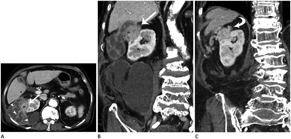
Fig. 1 Renal malakoplakia with wide retroperitoneal extension in a 78-year-old woman. Axial (A) and coronal (B) contrast enhanced CT scan shows a large multilocular cystic mass with irregular wall and septa (arrowheads) in right kidney and perirenal space. The mass extends into the right retroperitoneum, bare area and right psoas muscle. Focal enhancing solid portion is demonstrated in the right kidney (arrow). On follow-up CT scan (C) obtained after 4 months with antibiotic treatment, the complicated cystic lesion is markedly reduced. And the enhancing solid portion is also decreased in size (curved arrow).
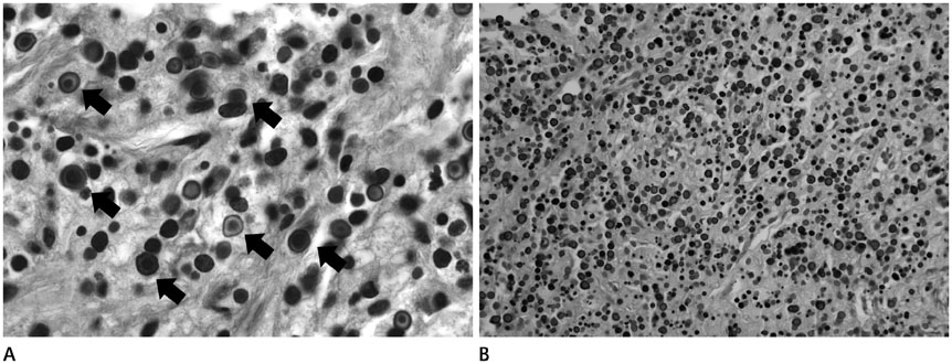
Fig. 2 Photomicrographs of malakoplakia. A. There are many scattered histiocytes (von Hansemann cells) containing intracytoplasmic lamellated basophilic inclusions, called Michaelis-Gutmann bodies (black arrows) (× 1000). B. A von Kossa stain highlights numerous Michaelis-Gutmann bodies (× 200).
Reference
-
1. Wielenberg AJ, Demos TC, Rangachari B, Turk T. Malacoplakia presenting as a solitary renal mass. AJR Am J Roentgenol. 2004; 183:1703–1705.2. Radin DR, Siskind B, Weiner S, Bernstein R, Ziprkowski M. Retroperitoneal malacoplakia. Urol Radiol. 1984; 6:218–220.3. Zimina OG, Rezun S, Armao D, Braga L, Semelka RC. Renal malacoplakia: demonstration by MR imaging. Magn Reson Imaging. 2002; 20:611–614.4. Kajbafzadeh A, Baharnoori M. Renal malakoplakia simulating neoplasm in a child: successful medical management. Urol J. 2004; 1:218–220.5. Yoon SY, Lee HJ, An JH, Kim SJ, Kim SW, Woo JH, et al. Renal parenchymal malakoplakia presenting with abscesses and hepatic extension misdiagnosed as a malignant tumor: a case report. Korean J Med. 2012; 82:764–768.6. Cury J, Coelho RF, Franco M, Srougi M. Renal parenchymal malacoplakia with pleural effusion. Clinics (Sao Paulo). 2007; 62:87–88.7. Dharmadhikari R, Crisp A. Sequential changes in sonographic appearances of childhood renal malakoplakia progressing to end-stage renal failure. J Ultrasound Med. 2006; 25:1219–1222.8. Kamishima T, Ito K, Awaya H, Mitchell DG. MR imaging of bilateral renal malacoplakia after liver transplantation. AJR Am J Roentgenol. 2000; 175:919–920.
- Full Text Links
-
- Actions
-
Cited
- CITED
-
- Close
- Share
-
- Similar articles
-
- Renal Malakoplakia with Secondary Hepatic Extension: A Case Report
- Pulmonary Malakoplakia Associated with Peripheral Cysts in an Immunocompetent Patient: A Case Report
- Renal Parenchymal Malakoplakia Presenting with Abscesses and Hepatic Extension Misdiagnosed as a Malignant Tumor: A Case Report
- Renal Parenchymal Malakoplakia with Acute Interstitial Nephritis Presented with Acute Kidney Injury
- Malakoplakia in a Renal Allograft: A case report
