Xanthogranulomatous Inflammation in Terminal Ileum Presenting as an Appendiceal Mass: Case Report and Review of the Literature
- Affiliations
-
- 1Department of Internal Medicine, Hanyang University Guri Hospital, Hanyang University College of Medicine, Guri, Korea. ycjeon@hanyang.ac.kr
- 2Department of Surgery, Hanyang University Guri Hospital, Hanyang University College of Medicine, Guri, Korea.
- 3Department of Pathology, Hanyang University Guri Hospital, Hanyang University College of Medicine, Guri, Korea.
- KMID: 1805305
- DOI: http://doi.org/10.5946/ce.2013.46.2.193
Abstract
- Xanthogranulomatous inflammation (XGI) is a rare benign inflammatory disease characterized by aggregation of lipid-laden foamy macrophages. This disease entity has been described in various organs but most commonly in the kidney and gallbladder. The occurrence of this disease in the lower gastrointestinal tract is extremely rare. Its clinical importance is that it can be misdiagnosed as an infiltrative cancer. In this case report, a 52-year-old male complained of right lower quadrant abdominal pain for a period of 3 months. Abdominal computed tomography revealed appendiceal mass and colonoscopy revealed multiple erythematous nodular lesions in the terminal ileum and appendiceal orifice, mimicking appendiceal cancer. Right hemicolectomy was done and the pathological specimen revealed XGI of the terminal ileum. To our knowledge, this is the first case of XGI in terminal ileum presenting as abdominal pain and the appendiceal mass on radiologic findings.
MeSH Terms
Figure
-
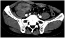
Fig. 1 Computed tomography (CT) findings. Abdominal CT images showed an approximately 5-cm appendiceal mass.

Fig. 2 Endoscopic findings. Colonoscopy showed multiple erythematous nodular lesions in (A) the terminal ileum and (B) appendiceal orifice.
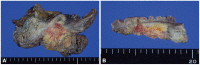
Fig. 3 Macroscopic findings. Cut section reveals (A) irregular golden-yellow mass-like lesion of the terminal ileum wall and mesentery (B) with a focus of mucosal lesion of the ileum.

Fig. 4 Microscopic findings. Histologic findings of xanthogranulomatous inflammation showing (A) nodular aggregation of lipid-laden foamy histiocytes (H&E stain, ×400), and (B) foreign body giant cell reaction surrounding food material (H&E stain, ×400), and (C) calcified needle-shaped foreign material (H&E stain, ×400).
Cited by 1 articles
-
A Case of Xanthogranulomatous Inflammation of Terminal Ileum Presenting as a Mass in a Woman with Severe Obesity
Hyung Ku Chon, Sang Wook Kim
Korean J Gastroenterol. 2016;67(5):277-281. doi: 10.4166/kjg.2016.67.5.277.
Reference
-
1. Cozzutto C, Carbone A. The xanthogranulomatous process. Xanthogranulomatous inflammation. Pathol Res Pract. 1988; 183:395–402. PMID: 3054826.2. Franco V, Aragona F, Genova G, Florena AM, Stella M, Campesi G. Xanthogranulomatous cholecystitis. Histopathological study and classification. Pathol Res Pract. 1990; 186:383–390. PMID: 2377572.3. Antonakopoulos GN, Chapple CR, Newman J, et al. Xanthogranulomatous pyelonephritis. A reappraisal and immunohistochemical study. Arch Pathol Lab Med. 1988; 112:275–281. PMID: 3345125.4. Oh YH, Seong SS, Jang KS, et al. Xanthogranulomatous inflammation presenting as a submucosal mass of the sigmoid colon. Pathol Int. 2005; 55:440–444. PMID: 15982221.
Article5. Maeda T, Shimada M, Matsumata T, et al. Xanthogranulomatous cholecystitis masquerading as gallbladder carcinoma. Am J Gastroenterol. 1994; 89:628–630. PMID: 8147372.6. Davis M, Whitley ME, Haque AK, Fenoglio-Preiser C, Waterman R. Xanthogranulomatous abscess of a mullerian duct remnant. A rare lesion of the rectum and anus. Dis Colon Rectum. 1986; 29:755–759. PMID: 3769695.7. Lo CY, Lorentz TG, Poon CS. Xanthogranulomatous inflammation of the sigmoid colon: a case report. Aust N Z J Surg. 1996; 66:643–644. PMID: 8859170.8. Anadol AZ, Gonul II, Tezel E. Xanthogranulomatous inflammation of the colon: a rare cause of cecal mass with bleeding. South Med J. 2009; 102:196–199. PMID: 19194269.9. Dhawan S, Jain D, Kalhan SK. Xanthogranulomatous inflammation of ascending colon with mucosal involvement: report of a first case. J Crohns Colitis. 2011; 5:245–248. PMID: 21575889.
Article10. Krishnani N, Dhingra S, Kapoor S, Pandey R. Cytopathologic diagnosis of xanthogranulomatous cholecystitis and coexistent lesions. A prospective study of 31 cases. Acta Cytol. 2007; 51:37–41. PMID: 17328493.
- Full Text Links
-
- Actions
-
Cited
- CITED
-
- Close
- Share
-
- Similar articles
-
- A Case of Xanthogranulomatous Inflammation of Terminal Ileum Presenting as a Mass in a Woman with Severe Obesity
- Appendiceal Diverticulosis Presented with Cecal Dicerticulitis
- A Case of Xanthogranulomatous Pyelonephritis Associated with Xanthogranulomatous Epididymoorchitis
- Hodgkin's sarcoma at the terminal ileum causing intussusception: a case report and review of the literature
- Xanthogranulomatous Cystitis
