Recent Advances in Image-enhanced Endoscopy
- Affiliations
-
- 1Division of Gastroenterology and Hepatology, Gastrointestinal Cancer Center, Soonchunhyang University Hospital, Seoul, Korea.
- 2Division of Gastroenterology, Department of Internal Medicine, College of Medicine, Kyung Hee University, Seoul, Korea. jyjang@khu.ac.kr
- 3Division of Gastroenterology, Department of Internal Medicine, Inha University Hospital, Incheon, Korea.
- KMID: 1805252
- DOI: http://doi.org/10.5946/ce.2011.44.2.65
Abstract
- The desire to better recognized such malignancies, which may be difficult to distinguish from inflammation or trauma, has accelerated the development of endoscopy with new optical technologies. Narrow-band imaging is a novel endoscopic technique that may enhance the accuracy of diagnosis using narrow-bandwidth filters in a red-green-blue sequential illumination system. Autofluorescence imaging is based on the detection of natural tissue fluorescence emitted by endogenous molecules. I-scan technology using a digital filter that modifies normal images through software functions, is the newly developed image-enhanced endoscopic technology from PENTAX. Flexible spectral imaging color enhancement enhances the visualization of mucosal structure and microcirculation by the selection of spectral transmittance with a dedicated wavelength. Confocal laser endomicroscopy images were collected with an argon beam with a scanning depth of 0 (epithelium) to 250 microm (lamina propria) and analyzed using the reflected light.
Keyword
MeSH Terms
Figure
-
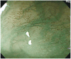
Fig. 1 The intra-epithelial papillary capillary loop (IPCL) image of normal esophageal mucosa in magnifying endoscopy using narrow-band imaging. Branching vessel which are located at the surface of muscularis mucosa are shown as a green vascular network. The IPCL is observed as a brown vessel which is positioned in the most superficial layer and is derived upright from the branching vessel.
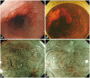
Fig. 2 Esophageal squamous cell carcinoma. (A) A depressed lesion with irregular nodularity and redness is noted at the mid esophagus. (B) With iodine staining, it is shown as an iodine-void area with a well-defined boundary. The change of intra-epithelial papillary capillary loop type V-3 and VN are observed in (C) the proximal margin and (D) center of the lesion. This lesion was diagnosed as SM2 cancer with lymphatic metastasis.
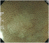
Fig. 3 Magnified endoscopic findings of light blue crests in the gastric antral mucosa. Light blue crest is clearly visualized as blue-white lines on the epithelial edge or surface by magnification with narrow-band imaging.
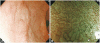
Fig. 4 Low grade dysplasia. (A) A slightly elevated discolored lesion is observed in the antrum. (B) White opaque substances along the surface of the lesion is observed by magnified endoscopy with narrow-band imaging.
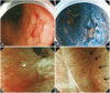
Fig. 5 Type 0-IIc early gastric cancer of well-differentiated adenocarcinoma. (A) A depressed lesion with central nodule is noted by white light endoscopy. (B) Chromoendoscopy. (C) Magnifying endoscopy with narrow-band imaging demonstrates loss of fine mucosal structure, loss of subepithelial capillary network and presence of an irregular microvascular pattern. (D) At the margin of the carcinoma, demarcation line is noted (arrows).
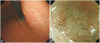
Fig. 6 Type 0-IIb early gastric cancer of signet ring cell carcinoma. (A) A flat pale mucosal lesion is noted on the body. (B) Magnifying endoscopy with narrow-band imaging findings of that pale mucosa show loss of the regular subepithelial capillary network pattern and corkscrew pattern.

Fig. 7 Type 0-IIc early gastric cancer of signet ring cell carcinoma. (A) A depressed pale mucosal lesion is noted on the high body. (B, C) Magnifying endoscopy with narrow-band imaging finding shows loss of the microsurface structure, corkscrew, and interrupted microvascular pattern. The vessels in cancerous lesions shows abnormal dilatation, abrupt in caliber and heterogeneity in shape. This lesion was diagnosed as SM1 cancer with lymphatic invasion.
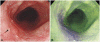
Fig. 8 Early esophageal cancer. (A) Conventinal endoscopic image shows flat lesion at left side (arrow). (B) Autofluorescence imaging shows that extent of the tumor become purple color change.
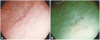
Fig. 9 I-scan image of depressed typed early gastric cancer. (A) Small depressed lesion is noted at antrum. (B) I-scan tone enhancement (TE)-g makes it more clear delineation of the tumor.

Fig. 10 Confocal laser endomicroscopy imaging of early esophageal cancer. (A) Normal esophageal mucosa shows regular patterns of squamous epithelium with normal intra-epithelial papillary capillary loop (IPCL) (arrow). (B) Tumor shows irregular patterns of squamous epithelium and dilated IPCL (arrow).

Fig. 11 Tumor margin delineation with confocal laser endomicroscopy (CLE). (A) Slightly depressed lesion is noted and previous forceps biopsy show signet ring cell adenocarcinoma. (B) It is not easily delineated for the exact tumor margin (red circle). (C) CLE help the delineation of the tumor margin with the determination of in vivo histology (normal mucosal surface; purple arrow in B). (D) Tumor mucosal surface (yellow arrow in B).
Cited by 4 articles
-
Increased Detection of Colorectal Polyps in Screening Colonoscopy Using High Definition i-SCAN Compared with Standard White Light
Woo Jung Kim, Sang Young Park, Iksoo Park, Wook Jin Lee, Jaechan Park, Nuri Chon, Tak Geun Oh, Kwang Hyun Kim
Clin Endosc. 2016;49(1):69-75. doi: 10.5946/ce.2016.49.1.69.Molecular Imaging for Theranostics in Gastroenterology: One Stone to Kill Two Birds
Kwang Hyun Ko, Chang-Il Kown, Jong Min Park, Hoo Geun Lee, Na Young Han, Ki Baik Hahm
Clin Endosc. 2014;47(5):383-388. doi: 10.5946/ce.2014.47.5.383.Image Quality Analysis of Various Gastrointestinal Endoscopes: Why Image Quality Is a Prerequisite for Proper Diagnostic and Therapeutic Endoscopy
Weon Jin Ko, Pyeong An, Kwang Hyun Ko, Ki Baik Hahm, Sung Pyo Hong, Joo Young Cho
Clin Endosc. 2015;48(5):374-379. doi: 10.5946/ce.2015.48.5.374.The Past, Present, and Future of Image-Enhanced Endoscopy
Jae-Young Jang
Clin Endosc. 2015;48(6):466-475. doi: 10.5946/ce.2015.48.6.466.
Reference
-
1. Tajiri H, Matsuda K, Fujisaki J. What can we see with the endoscope? Present status and future perspectives. Dig Endosc. 2002; 14:131–137.
Article2. Kara MA, Peters FP, Rosmolen WD, et al. High-resolution endoscopy plus chromoendoscopy or narrow-band imaging in Barrett's esophagus: a prospective randomized crossover study. Endoscopy. 2005; 37:929–936. PMID: 16189764.
Article3. Yasushi S, Manabu M, Hisao T, Atsushi O, Shigeaki Y. Optical/digital chromoendoscopy during colonoscopy using narrow-band imaging system. Dig Endosc. 2005; 17(Suppl 1):S43–S48.4. Muto M, Katada C, Sano Y, Yoshida S. Narrow band imaging: a new diagnostic approach to visualize angiogenesis in superficial neoplasia. Clin Gastroenterol Hepatol. 2005; 3(7 Suppl 1):S16–S20. PMID: 16012987.
Article5. Singh S, Sharma P. Magnification endoscopy in the upper GI tract. Dig Endosc. 2005; 17(Suppl 1):S17–S19.
Article6. Sharma P, Weston AP, Topalovski M, Cherian R, Bhattacharyya A, Sampliner RE. Magnification chromoendoscopy for the detection of intestinal metaplasia and dysplasia in Barrett's oesophagus. Gut. 2003; 52:24–27. PMID: 12477754.
Article7. Hamamoto Y, Endo T, Nosho K, Arimura Y, Sato M, Imai K. Usefulness of narrow-band imaging endoscopy for diagnosis of Barrett's esophagus. J Gastroenterol. 2004; 39:14–20. PMID: 14767729.
Article8. Inoue H, Honda T, Yoshida T, et al. Ultra-high magnification endoscopy of the normal esophageal mucosa. Dig Endosc. 1996; 8:134–138.
Article9. Inoue H, Honda T, Nagai K, et al. Ultra-high magnification endoscopic observation of carcinoma in situ of the esophagus. Dig Endosc. 1997; 9:16–18.
Article10. Inoue H. Magnification endoscopy in the esophagus and stomach. Dig Endosc. 2001; 13(Suppl 1):S40–S41.
Article11. Yoshida T, Inoue H, Usui S, Satodate H, Fukami N, Kudo SE. Narrow-band imaging system with magnifying endoscopy for superficial esophageal lesions. Gastrointest Endosc. 2004; 59:288–295. PMID: 14745410.
Article12. Capelle LG, Haringsma J, de Vries AC, et al. Narrow band imaging for the detection of gastric intestinal metaplasia and dysplasia during surveillance endoscopy. Dig Dis Sci. 2010; 55:3442–3448. PMID: 20393882.
Article13. Anagnostopoulos GK, Yao K, Kaye P, et al. High-resolution magnification endoscopy can reliably identify normal gastric mucosa, Helicobacter pylori-associated gastritis, and gastric atrophy. Endoscopy. 2007; 39:202–207. PMID: 17273960.
Article14. Uedo N, Ishihara R, Iishi H, et al. A new method of diagnosing gastric intestinal metaplasia: narrow-band imaging with magnifying endoscopy. Endoscopy. 2006; 38:819–824. PMID: 17001572.
Article15. Yao K, Iwashita A, Tanabe H, et al. White opaque substance within superficial elevated gastric neoplasia as visualized by magnification endoscopy with narrow-band imaging: a new optical sign for differentiating between adenoma and carcinoma. Gastrointest Endosc. 2008; 68:574–580. PMID: 18656862.
Article16. Yao K, Yao T, Iwashita A. Determining the horizontal extent of early gastric carcinoma: two modern techniques based on differences in the mucosal microvascular architecture and density between carcinomatous and non-carcinomatous mucosa. Dig Endosc. 2002; 14(Suppl 1):S83–S87.
Article17. Nakayoshi T, Tajiri H, Matsuda K, Kaise M, Ikegami M, Sasaki H. Magnifying endoscopy combined with narrow band imaging system for early gastric cancer: correlation of vascular pattern with histopathology (including video). Endoscopy. 2004; 36:1080–1084. PMID: 15578298.
Article18. Yagi K, Nakamura A, Sekine A, Hajime U. Magnifying endoscopy with narrow band imaging for early differentiated gastric adenocarcinoma. Dig Endosc. 2008; 20:115–122.
Article19. Kadowaki S, Tanaka K, Toyoda H, et al. Ease of early gastric cancer demarcation recognition: a comparison of four magnifying endoscopy methods. J Gastroenterol Hepatol. 2009; 24:1625–1630. PMID: 19788603.
Article20. Kiyotoki S, Nishikawa J, Satake M, et al. Usefulness of magnifying endoscopy with narrow-band imaging for determining gastric tumor margin. J Gastroenterol Hepatol. 2010; 25:1636–1641. PMID: 20880172.
Article21. Ezoe Y, Muto M, Horimatsu T, et al. Magnifying narrow-band imaging versus magnifying white-light imaging for the differential diagnosis of gastric small depressive lesions: a prospective study. Gastrointest Endosc. 2010; 71:477–484. PMID: 20189506.
Article22. Kato M, Kaise M, Yonezawa J, et al. Magnifying endoscopy with narrow-band imaging achieves superior accuracy in the differential diagnosis of superficial gastric lesions identified with white-light endoscopy: a prospective study. Gastrointest Endosc. 2010; 72:523–529. PMID: 20598685.
Article23. Cho JY, Hong SJ. Autofluorescence imaging: as a new method for predicting metachronous gastric cancer. J Gastroenterol Hepatol. 2010; 25:1814–1815. PMID: 21091990.
Article24. Otani A, Amano Y, Koshino K, et al. Is autofluorescence imaging endoscopy useful for determining the depth of invasion in gastric cancer? Digestion. 2010; 81:96–103. PMID: 20068309.
Article25. Nakamura M, Tahara T, Shibata T, et al. Diagnostic efficacy of autofluorescence and reflectance imaging endoscopy for lateral extension of early gastric cancers. Gastrointest Endosc. 2009; 70:599. PMID: 19699985.
Article26. Kim WJ, Cho JY, Jeong SW, et al. Comparison of autofluorescence imaging endoscopic findings with pathologic findings after endoscopic submucosal dissection of gastric neoplasms. Gut Liver. 2008; 2:186–192. PMID: 20485645.
Article27. Kato M, Uedo N, Ishihara R, et al. Analysis of the color patterns of early gastric cancer using an autofluorescence imaging video endoscopy system. Gastric Cancer. 2009; 12:219–224. PMID: 20047127.
Article28. Kara MA, Peters FP, Fockens P, ten Kate FJ, Bergman JJ. Endoscopic video-autofluorescence imaging followed by narrow band imaging for detecting early neoplasia in Barrett's esophagus. Gastrointest Endosc. 2006; 64:176–185. PMID: 16860064.
Article29. Ohkawa A, Miwa H, Namihisa A, et al. Diagnostic performance of light-induced fluorescence endoscopy for gastric neoplasms. Endoscopy. 2004; 36:515–521. PMID: 15202048.30. Ignjatovic A, East JE, Guenther T, et al. What is the most reliable imaging modality for small colonic polyp characterization? Study of white-light, autofluorescence, and narrow-band imaging. Endoscopy. 2011; 43:94–99. PMID: 21271465.
Article31. Lee BI. EPK-i endoscopy. Korean J Gastrointest Endosc. 2009; 39(Suppl 1):184–186.32. Kodashima S, Fujishiro M. Novel image-enhanced endoscopy with i-scan technology. World J Gastroenterol. 2010; 16:1043–1049. PMID: 20205272.
Article33. Hoffman A, Kagel C, Goetz M, et al. Recognition and characterization of small colonic neoplasia with high-definition colonoscopy using i-scan is as precise as chromoendoscopy. Dig Liver Dis. 2010; 42:45–50. PMID: 19473893.
Article34. Hoffman A, Sar F, Goetz M, et al. High definition plus colonoscopy combined with i-scan is superior in the detection and characterization of colorectal neoplasias compared to standard video colonoscopy: a prospective randomized trial. Gastrointest Endosc. 2009; 69:AB131–AB132.35. Hong SW, Cho WY, Cho JY, et al. Comparison between i scan and ch-romoscopy for delineation of the margin in early gastric cancer. Endoscopy. 2011; Forthcoming.36. Yoshizawa M, Osawa H, Yamamoto H, et al. Diagnosis of elevated-type early gastric cancers by the optimal band imaging system. Gastrointest Endosc. 2009; 69:19–28. PMID: 19111685.
Article37. Osawa H, Yoshizawa M, Yamamoto H, et al. Optimal band imaging system can facilitate detection of changes in depressed-type early gastric cancer. Gastrointest Endosc. 2008; 67:226–234. PMID: 18061596.
Article38. Kiesslich R, Burg J, Vieth M, et al. Confocal laser endoscopy for diagnosing intraepithelial neoplasias and colorectal cancer in vivo. Gastroenterology. 2004; 127:706–713. PMID: 15362025.
Article39. Kiesslich R, Gossner L, Goetz M, et al. In vivo histology of Barrett's esophagus and associated neoplasia by confocal laser endomicroscopy. Clin Gastroenterol Hepatol. 2006; 4:979–987. PMID: 16843068.
Article40. Liu H, Li YQ, Yu T, et al. Confocal endomicroscopy for in vivo detection of microvascular architecture in normal and malignant lesions of upper gastrointestinal tract. J Gastroenterol Hepatol. 2008; 23:56–61. PMID: 18028347.
Article41. Liu H, Li YQ, Yu T, et al. Confocal laser endomicroscopy for superficial esophageal squamous cell carcinoma. Endoscopy. 2009; 41:99–106. PMID: 19214886.
Article42. Jeon SR, Cho WY, Jin SY, Cheon YK, Choi SR, Cho JY. Optical biopsies by confocal endomicroscopy prevent additive endoscopic biopsies before endoscopic submucosal dissection in gastric epithelial neoplasias: a prospective, comparative study. Gastrointest Endosc. 2011; 74:772–780. PMID: 21802680.
Article
- Full Text Links
-
- Actions
-
Cited
- CITED
-
- Close
- Share
-
- Similar articles
-
- Is Image-Enhanced Endoscopy Useful for the Diagnosis and Treatment of Gastrointestinal Tumor?
- Role of Image-Enhanced Endoscopy in Pancreatobiliary Diseases
- Preparation of image databases for artificial intelligence algorithm development in gastrointestinal endoscopy
- Current status of image-enhanced endoscopy in inflammatory bowel disease
- The Past, Present, and Future of Image-Enhanced Endoscopy
