The Past, Present, and Future of Image-Enhanced Endoscopy
- Affiliations
-
- 1Division of Gastroenterology, Department of Internal Medicine, Kyung Hee University School of Medicine, Seoul, Korea. jyjang@khu.ac.kr
- KMID: 2380402
- DOI: http://doi.org/10.5946/ce.2015.48.6.466
Abstract
- Despite the remarkable progress recently made to enhance the resolution of white-light endoscopy, detection, and diagnosis of premalignant lesions, such as adenomas and subtle early-stage cancers, remains a great challenge. As for example, although chromoendoscopy, such as endoscopy using indigo carmine, is useful for the early diagnosis of subtle lesions, the technique presents various disadvantages ranging from the time required for spray application of the dye and suctioning of excess dye to the increased difficulty in identifying lesions in the presence of severe inflammation and obstruction of visual field due to the pooling of solution in depressed-type lesions. To overcome these diagnostic problems associated with chromoendoscopy, research has focused on the development of endoscopes based on new optical technologies. Several types of image-enhanced endoscopy methods have recently been presented. In particular, image-enhanced endoscopy has emerged as a new paradigm for the diagnosis of gastrointestinal disorders. Image-enhanced endoscopes provide high-contrast images of lesions by means of optical or electronic technologies, including the contrast enhancement of the mucosal surface and of blood vessels. Chromoendoscopy, narrow-band imaging, i-SCAN, and flexible spectral imaging color enhancement are representative examples of image-enhanced endoscopy discussed in this paper.
Keyword
MeSH Terms
Figure
-
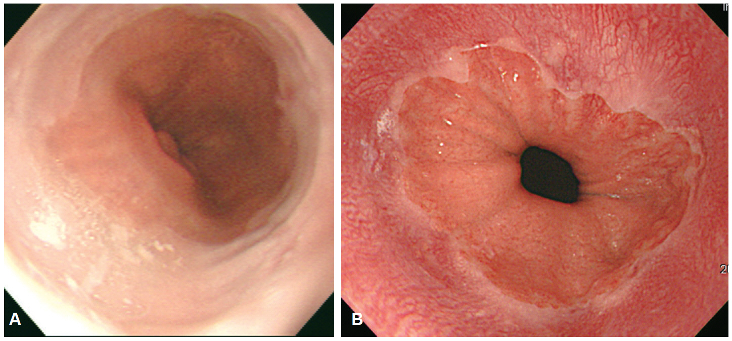
Fig. 1. The resolution of endoscopic image. (A) Standard definition (SD) endoscopic image of gastroesophageal (GE) junction, (B) high definition endoscopic image of GE junction can be observed more clearly compared to the SD endoscopic image.
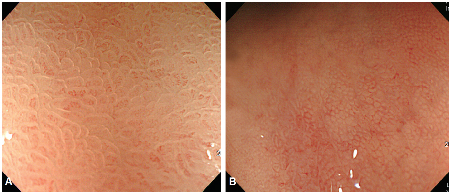
Fig. 2. Magnifying endoscopic finding of normal gastric mucosa. (A) In the gastric antrum, microvascular architecture is a coil-shaped. The pits demonstrate a linear or reticular patterns. (B) In the gastric body, microvascular architecture is a honeycomb shaped, and the pits demonstrate a round or oval shape.
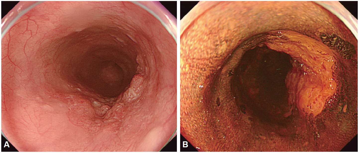
Fig. 3. Esophageal squamous cell carcinoma. (A) A depressed lesion with irregular nodularity and redness is noted at the mid esophagus. (B) With iodine staining, it is shown as an iodine-void area with a well-defined boundary.
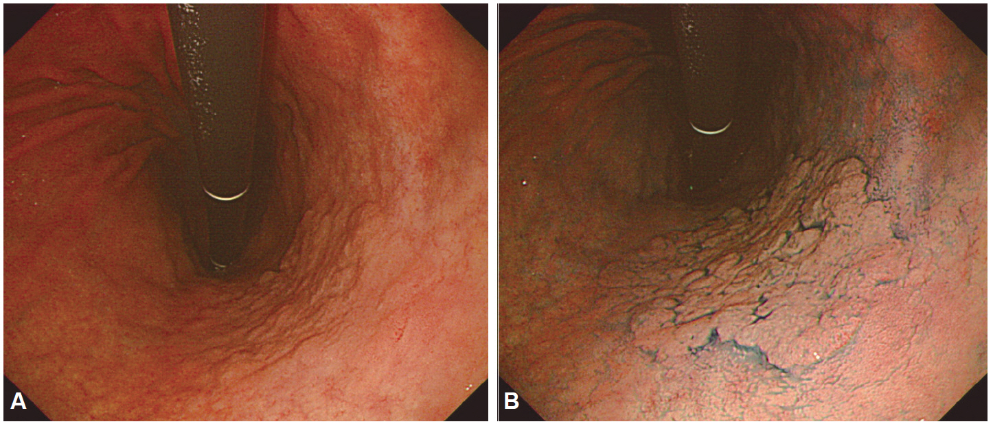
Fig. 4. Early gastric cancer. (A) Indistinct margined gastric cancer due to marked atrophy and intestinal metaplasia around the lesions are often observed. (B) Indigo carmine chromoendoscopy delineated the lesion more clearly.
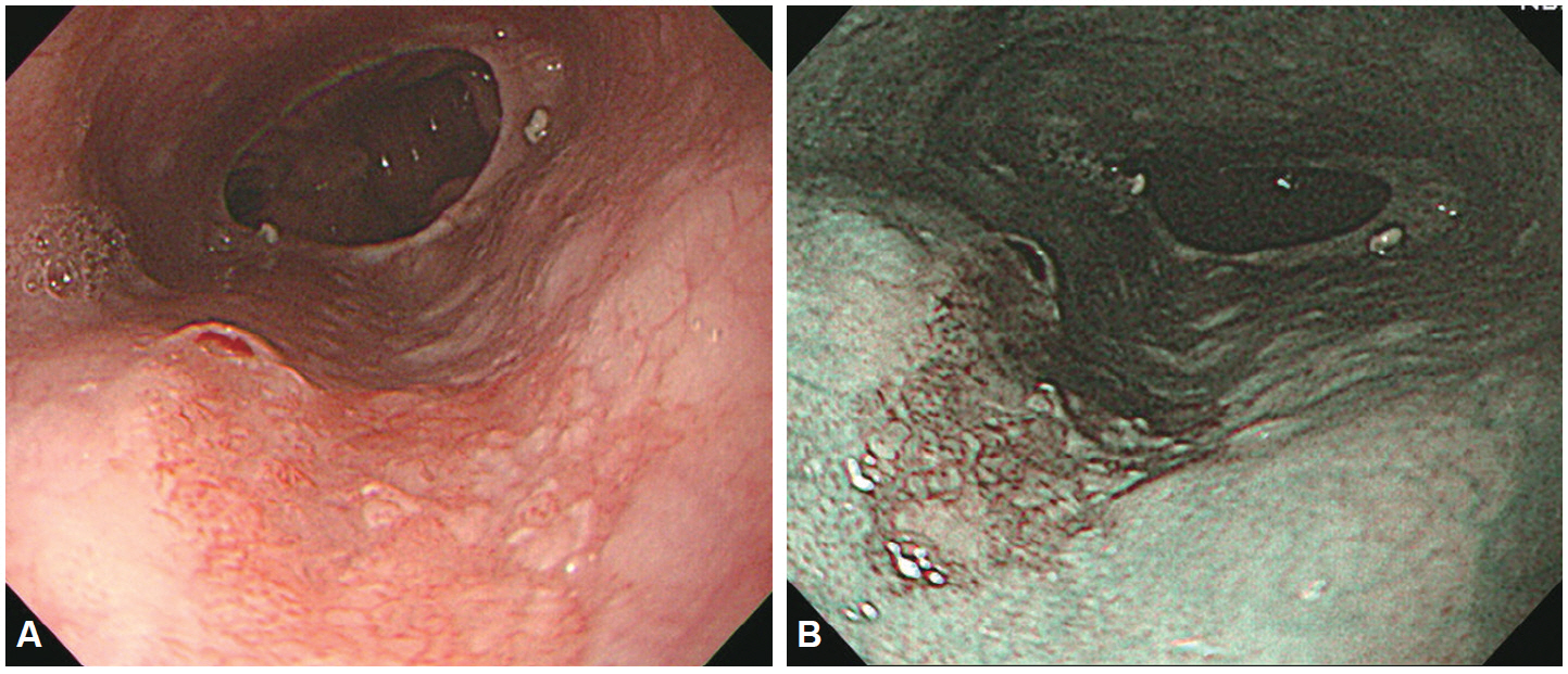
Fig. 5. Esophageal squamous cell carcinoma. (A) A depressed lesion with irregular nodularity and redness is noted at the lower esophagus. (B) In narrow-band imaging mode, is seen as dark brown lesion due to its copious vascularity.
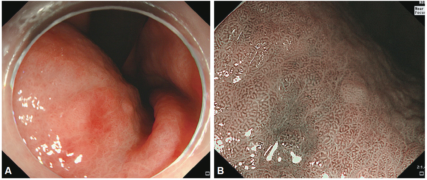
Fig. 6. Early gastric cancer. (A) About 0.6-cm sized slightly depressed lesion was noted at the lesser curvature side of mid body in white light imaging. (B) Well delineated margin, loss of pit, and irregular microvessels were noted in near focus.
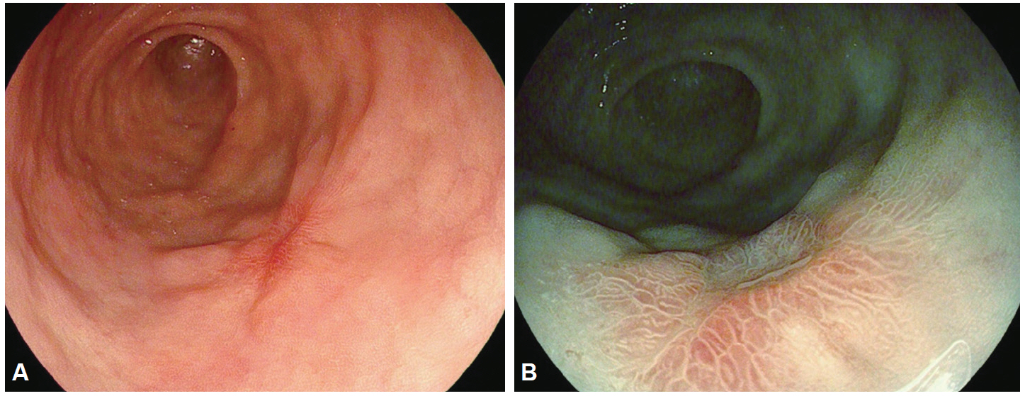
Fig. 7. i-SCAN image of depressed typed early gastric cancer. (A) Small depressed lesion is noted at antrum. (B) i-SCAN TE-g makes it more clear delineation of the tumor.
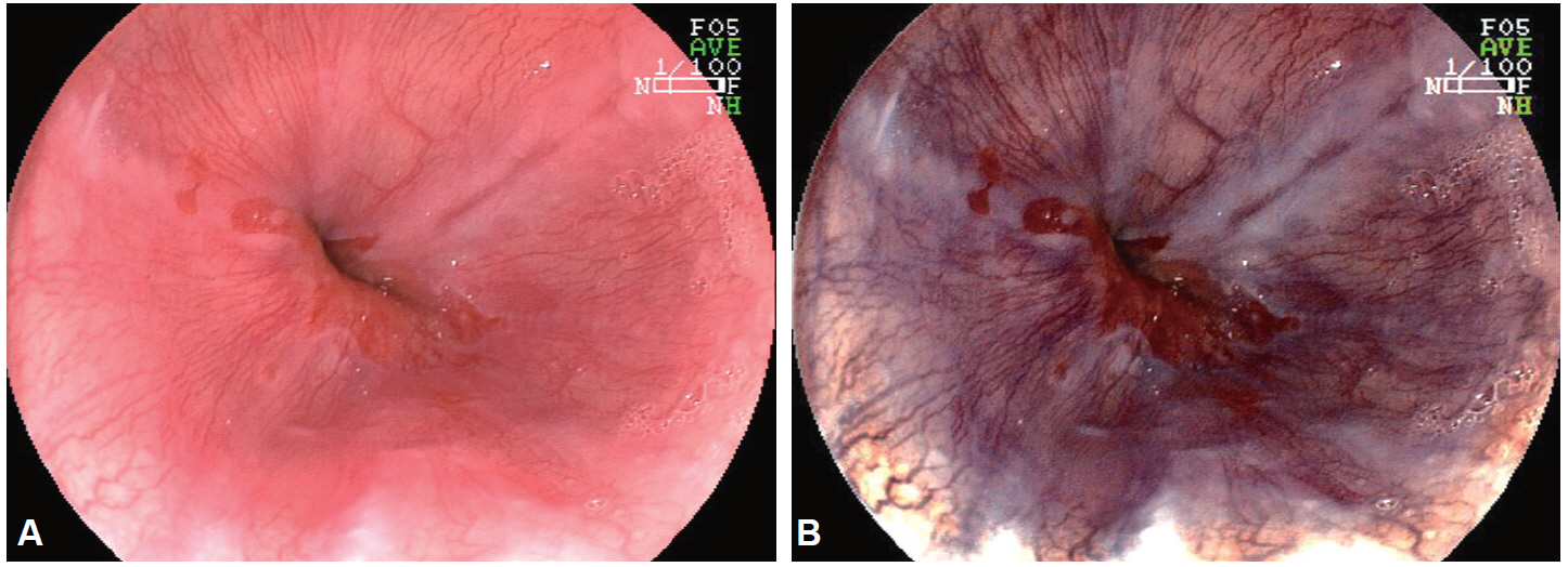
Fig. 8. Reflux esophagitis. (A) Gastroesophageal junction and a mucosal break are noted in white light imaging. (B) Flexible spectral imaging color enhancement imaging enhanced the visualization of mucosal structure and vessels.
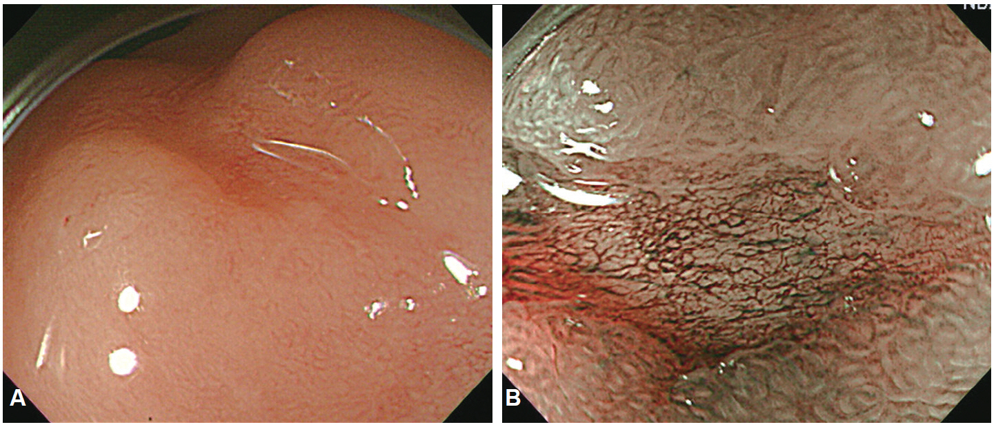
Fig. 9. Early gastric cancer. (A) A depressed lesion was noted at the antrum. (B) Magnifying narrow-band imaging demonstrated demarcation line, loss of fine mucosal structure, and presence of an irregular microvascular pattern. The lesion was suspected to mucosal well differentiated gastric adenocarcinoma. Endoscopic submucosal dissection was performed.
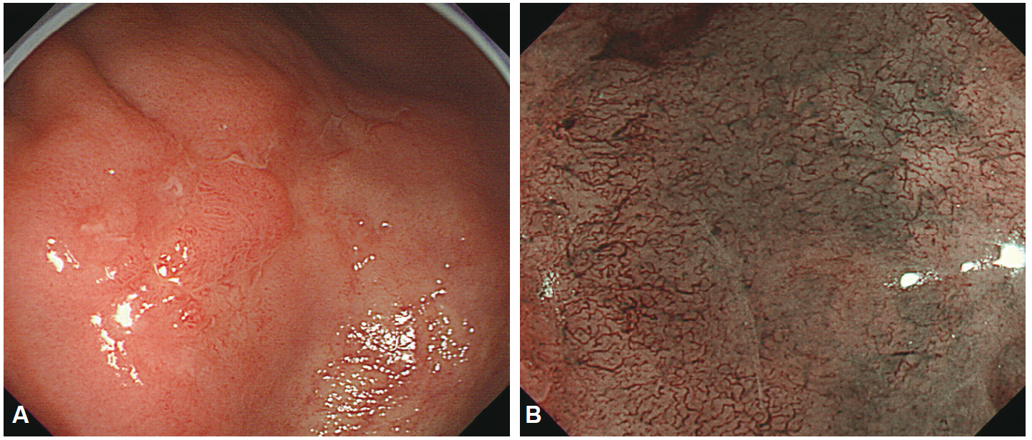
Fig. 10. Early gastric cancer. (A) Ill defined, pale mucosal lesion with central nodule is noted on the antrum. (B) Magnifying narrow-band imaging findings of that pale mucosa showed loss of the regular microvascular pattern and corkscrew pattern. The lesion was suspected to poorly differentiated gastric adenocarcinoma. Gastrectomy with lymph node dissection was performed.
Cited by 3 articles
-
Application of Current Image-Enhanced Endoscopy in Gastric Diseases
Wansik Lee
Clin Endosc. 2021;54(4):477-487. doi: 10.5946/ce.2021.160.The Role of Dual Red Imaging in Gastric Endoscopic Submucosal Dissection
In Kyung Yoo, Joo Young Cho
Clin Endosc. 2020;53(1):1-2. doi: 10.5946/ce.2020.018.Usefulness of Narrow-Band Imaging in Endoscopic Submucosal Dissection of the Stomach
Jung-Wook Kim
Clin Endosc. 2018;51(6):527-533. doi: 10.5946/ce.2018.186.
Reference
-
1. Subramanian V, Ragunath K. Advanced endoscopic imaging: a review of commercially available technologies. Clin Gastroenterol Hepatol. 2014; 12:368.e1–376.e1.
Article2. ASGE Technology Committee, Kwon RS, Adler DG, et al. High-resolution and high-magnification endoscopes. Gastrointest Endosc. 2009; 69(3 Pt 1):399–407.
Article3. Bruno MJ. Magnification endoscopy, high resolution endoscopy, and chromoscopy; towards a better optical diagnosis. Gut. 2003; 52 Suppl 4:iv7–iv11.
Article4. Peitz U, Malfertheiner P. Chromoendoscopy: from a research tool to clinical progress. Dig Dis. 2002; 20:111–119.
Article5. Woolf GM, Riddell RH, Irvine EJ, Hunt RH. A study to examine agreement between endoscopy and histology for the diagnosis of columnar lined (Barrett’s) esophagus. Gastrointest Endosc. 1989; 35:541–544.
Article6. Inoue H, Rey JF, Lightdale C. Lugol chromoendoscopy for esophageal squamous cell cancer. Endoscopy. 2001; 33:75–79.7. Kida M, Kobayashi K, Saigenji K. Routine chromoendoscopy for gastrointestinal diseases: indications revised. Endoscopy. 2003; 35:590–596.
Article8. Canto MI, Setrakian S, Petras RE, Blades E, Chak A, Sivak MV Jr. Methylene blue selectively stains intestinal metaplasia in Barrett’s esophagus. Gastrointest Endosc. 1996; 44:1–7.
Article9. Stevens PD, Lightdale CJ, Green PH, Siegel LM, Garcia-Carrasquillo RJ, Rotterdam H. Combined magnification endoscopy with chromoendoscopy for the evaluation of Barrett’s esophagus. Gastrointest Endosc. 1994; 40:747–749.
Article10. Siegel LM, Stevens PD, Lightdale CJ, et al. Combined magnification endoscopy with chromoendoscopy in the evaluation of patients with suspected malabsorption. Gastrointest Endosc. 1997; 46:226–230.
Article11. Axelrad AM, Fleischer DE, Geller AJ, et al. High-resolution chromoendoscopy for the diagnosis of diminutive colon polyps: implications for colon cancer screening. Gastroenterology. 1996; 110:1253–1258.
Article12. Sano Y, Muto M, Tajiri H, Ohtsu A, Yoshida S. Optical/digital chromoendoscopy during colonoscopy using narrow-band imaging system. Dig Endosc. 2005; 17 Suppl 1:S43–S48.
Article13. Muto M, Katada C, Sano Y, Yoshida S. Narrow band imaging: a new diagnostic approach to visualize angiogenesis in superficial neoplasia. Clin Gastroenterol Hepatol. 2005; 3(7 Suppl 1):S16–S20.
Article14. Cho WY, Jang JY, Lee DH; Endoscopic Technology and Investigation Study Group. Recent advances in image-enhanced endoscopy. Clin Endosc. 2011; 44:65–75.
Article15. Tajiri H, Matsuda K, Fujisaki J. What can we see with the endoscope? Present status and future perspectives. Dig Endosc. 2002; 14:131–137.
Article16. Kodashima S, Fujishiro M. Novel image-enhanced endoscopy with i-scan technology. World J Gastroenterol. 2010; 16:1043–1049.
Article17. Lee BI. EPK-i Endoscopy. Korean J Gastrointest Endosc. 2009; 39(Suppl 1):184–186.18. Yoshizawa M, Osawa H, Yamamoto H, et al. Diagnosis of elevated-type early gastric cancers by the optimal band imaging system. Gastrointest Endosc. 2009; 69:19–28.
Article19. Osawa H, Yoshizawa M, Yamamoto H, et al. Optimal band imaging system can facilitate detection of changes in depressed-type early gastric cancer. Gastrointest Endosc. 2008; 67:226–234.
Article20. ASGE Technology Committee, Manfredi MA, Abu Dayyeh BK, et al. Electronic chromoendoscopy. Gastrointest Endosc. 2015; 81:249–261.
Article21. Osawa H, Yamamoto H. Present and future status of flexible spectral imaging color enhancement and blue laser imaging technology. Dig Endosc. 2014; 26 Suppl 1:105–115.
Article
- Full Text Links
-
- Actions
-
Cited
- CITED
-
- Close
- Share
-
- Similar articles
-
- Is Image-Enhanced Endoscopy Useful for the Diagnosis and Treatment of Gastrointestinal Tumor?
- Past, Present, and Future of the Korea-Japan Joint Symposium on Gastrointestinal Endoscopy
- Application of Current Image-Enhanced Endoscopy in Gastric Diseases
- Quality Improvement of Gastrointestinal Endoscopy in Korea: Past, Present, and Future
- Role of Image-Enhanced Endoscopy in Pancreatobiliary Diseases
