Intrathyroidal Parathyroid Carcinoma: A Case Report
- Affiliations
-
- 1Department of Radiology, Hallym University College of Medicine, Dongtan Sacred Heart Hospital, Hwaseong, Korea.
- 2Department of Nuclear Medicine, Hallym University College of Medicine, Dongtan Sacred Heart Hospital, Hwaseong, Korea. youme@hallym.or.kr
- 3Department of Pathology, Hallym University College of Medicine, Dongtan Sacred Heart Hospital, Hwaseong, Korea.
- KMID: 1793887
- DOI: http://doi.org/10.3348/jksr.2015.72.5.319
Abstract
- Parathyroid carcinoma is an uncommon malignancy and a rare cause of primary hyperparathyroidism. Intrathyroidal parathyroid carcinoma is even rarer and only few cases have been reported previously. A 33-year-old woman presented with hypercalcemia. CT scan revealed a 5-cm sized intrathyroid nodule with a positive beak sign on the surface in contact with the thyroid gland. The patient underwent total thyroidectomy, and the histopathologic examination confirmed the diagnosis of parathyroid carcinoma. We report a case of intrathyroidal parathyroid carcinoma with brief literature review.
MeSH Terms
Figure
-
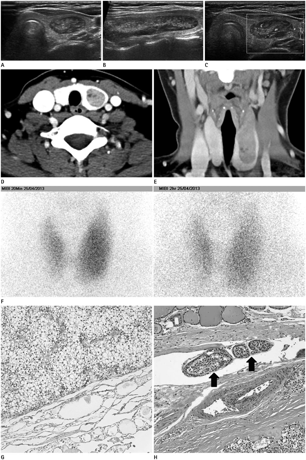
Fig. 1 A 33-year-old woman with intrathyroidal parathyroid carcinoma. Axial (A) and longitudinal (B) ultrasonogram shows a well marginated mixed echoic mass in left thyroid. On color Doppler ultrasonogram (C), marked increased vascularity is noted in left thyroid mass. Axial enhanced CT (D) reveals a well-marginated, heterogeneous enhancing mass in left thyroid gland. Coronal enhanced CT (E) shows an elongated left thyroid mass with a positive beak sign on the surface in contact with the thyroid gland. Parathyroid technetium-99m methoxyisobutylisonitrile scans (F). (Left) A 20-minute image shows diffuse even uptake by the left thyroid mass similar to that of the adjacent normal thyroid parenchyma. (Right) On a 2-hour delayed image, diffuse washout is noted in the both thyroid glands and left thyroid mass without definite focal abnormal hot uptake. Immunohistochemical staining (× 200) (G) for antiparathyroid hormone reveals strong parathyroid hormone expression in the resected tumor (upper portion). On histopathologic examination (H&E, × 200) (H), tumor cells (arrows) are found in the vascular space suggesting vascular invasion.
Cited by 1 articles
-
Intrathyroidal Parathyroid Carcinoma in Chronic Kidney Disease: A Case Report and Review of Literature
Moo Keon Kim, Chang Myeon Song, Kyung Tae, Yong Bae Ji
Korean J Otorhinolaryngol-Head Neck Surg. 2019;62(12):740-746. doi: 10.3342/kjorl-hns.2019.00409.
Reference
-
1. Koea JB, Shaw JH. Parathyroid cancer: biology and management. Surg Oncol. 1999; 8:155–165.2. Vila Duckworth L, Winter WE, Vaysberg M, Moran CA, Al-Quran SZ. Intrathyroidal parathyroid carcinoma: report of an unusual case and review of the literature. Case Rep Pathol. 2013; 2013:198643.3. Stewart AK, Bland KI, McGinnis LS Jr, Morrow M, Eyre HJ. Clinical highlights from the National Cancer Data Base, 2000. CA Cancer J Clin. 2000; 50:171–183.4. Moran CA, Suster S. Primary parathyroid tumors of the mediastinum: a clinicopathologic and immunohistochemical study of 17 cases. Am J Clin Pathol. 2005; 124:749–754.5. Castillo L, Poissonnet G, Haddad A, Guevara N, Santini J, Demard F. [Parathyroid carcinoma: diagnosis and treatment]. Rev Laryngol Otol Rhinol (Bord). 2000; 121:169–173.6. Chang YJ, Mittal V, Remine S, Manyam H, Sabir M, Richardson T, et al. Correlation between clinical and histological findings in parathyroid tumors suspicious for carcinoma. Am Surg. 2006; 72:419–426.7. Patel SB, Shah SR, Goswami KG, Patel HB. Pictorial essays: ultrasound features of thyroid and parathyroid lesions. Indian J Radiol Imaging. 2005; 15:211–216.8. Mazeh H, Kouniavsky G, Schneider DF, Makris KI, Sippel RS, Dackiw AP, et al. Intrathyroidal parathyroid glands: small, but mighty (a Napoleon phenomenon). Surgery. 2012; 152:1193–1200.9. Kitapçi MT, Tastekin G, Turgut M, Caner B, Kars A, Barista I, et al. Preoperative localization of parathyroid carcinoma using Tc-99m MIBI. Clin Nucl Med. 1993; 18:217–219.10. Ruan M, Shen Y, Zhang H, Li M, Chen L. Bone metastasis from parathyroid carcinoma non-avid for 99mTc-MIBI, 99mTc-MDP, and 18F-FDG. J Nucl Med Radiat Ther. 2014; 5:1–3. http://dx.doi.org/10.4172/2155-9619.1000165.
- Full Text Links
-
- Actions
-
Cited
- CITED
-
- Close
- Share
-
- Similar articles
-
- Intrathyroidal parathyroid carcinoma: a case report and literature review
- A Case of Hyperparathyroidism Caused by Intrathyroidal Parathyroid Adenoma
- A Case of Intrathyroidal Parathyroid Carcinoma Accompanied by Contralateral Parathyroid Hyperplasia
- A Case of Spontaneous Extracapsular Hemorrhage of Intrathyroidal Parathyroid Adenoma
- Identification of Intrathyroidal Parathyroid Gland Using Near-Infrared Autofluorescence and Autotransplantation: Report of Two Cases
