Korean J Ophthalmol.
2014 Apr;28(2):138-149. 10.3341/kjo.2014.28.2.138.
Reproducibility of Peripapillary Retinal Nerve Fiber Layer Thickness Measured by Spectral Domain Optical Coherence Tomography in Pseudophakic Eyes
- Affiliations
-
- 1Siloam Eye Hospital, Seoul, Korea. jhkim32@hanmail.net
- 2Institute of Vision Research, Department of Ophthalmology, Yonsei University College of Medicine, Seoul, Korea.
- KMID: 1792107
- DOI: http://doi.org/10.3341/kjo.2014.28.2.138
Abstract
- PURPOSE
To assess the reproducibility of circumpapillary retinal nerve fiber layer (cpRNFL) thickness measurement (measurement agreement) and its color-coded classification (classification agreement) by Cirrus spectral domain optical coherence tomography (OCT) in pseudophakic eyes.
METHODS
Two-hundred five participants having glaucoma or glaucoma suspected eyes underwent two repeated Cirrus OCT scans to measure cpRNFL thickness (optic disc cube 200 x 200). After classifying participants into three different groups according to their lens status (clear media, cataract, and pseudophakic), values of intra-class coefficient (ICC), coefficient of variance, and test-retest variability were compared between groups for average retinal nerve fiber layer (RNFL) thicknesses and that corresponding to four quadrant maps. Linear weighted kappa coefficients were calculated as indicators of agreement of color code classification in each group.
RESULTS
ICC values were all excellent (generally defined as 0.75 to 1.00) for the average and quadrant RNFL thicknesses in all three groups. ICC values of the clear media group tended to be higher than those in the cataract and pseudophakic groups for all quadrants and average thickness. Especially in the superior and nasal quadrants, the ICC value of the cataract group was significantly lower than that of the clear media and pseudophakic groups. For average RNFL thickness, classification agreement (kappa) in three groups did not show a statistically significant difference. For quadrant maps, classification agreement (kappa) in the clear media group was higher than those in the other two groups.
CONCLUSIONS
Agreement of cpRNFL measurement and its color code classification between two repeated Cirrus OCT scans in pseudophakic eyes was as good as that in eyes with clear crystalline lens. More studies are required to ascertain the effect of lens status on the reproducibility of Cirrus OCT according to different stages of glaucoma patients.
MeSH Terms
-
Aged
Cataract/complications
Cataract Extraction
Female
Glaucoma/complications/*pathology
Humans
Lens, Crystalline/cytology/pathology
Male
Middle Aged
Nerve Fibers/pathology
Optic Disk/pathology
Pseudophakia/complications
Reproducibility of Results
Retinal Ganglion Cells/*pathology
Tomography, Optical Coherence/*methods/*standards
Figure
-
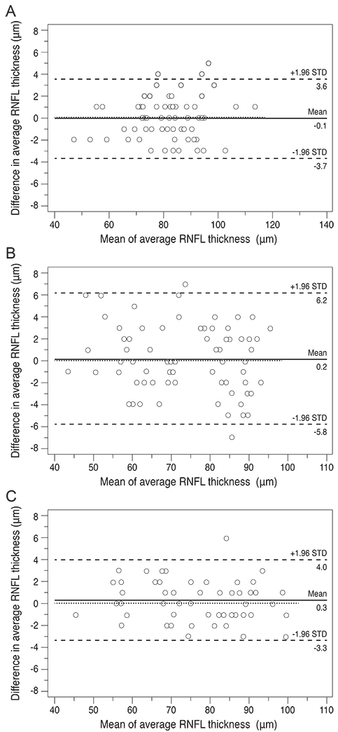
Fig. 1 Bland-Altman plot analysis of the average retinal nerve fiber layer (RNFL) thickness measured by two repeated Cirrus optical coherence tomography scans. Note the greater variation between two repeated average RNFL thickness measurements in the cataract group than in the other groups. (A) Clear media. (B) Cataract. (C) Pseudophakic. STD = standard deviation.
Reference
-
1. Carpineto P, Nubile M, Agnifili L, et al. Reproducibility and repeatability of Cirrus HD-OCT peripapillary retinal nerve fibre layer thickness measurements in young normal subjects. Ophthalmologica. 2012; 227:139–145.2. Cremasco F, Massa G, Goncalves Vidotti V, et al. Intrasession, intersession, and interexaminer variabilities of retinal nerve fiber layer measurements with spectral-domain OCT. Eur J Ophthalmol. 2011; 21:264–270.3. Pareja-Esteban J, Teus-Guezala MA, Drake-Casanova P, Dapena-Sevilla I. Retinal nerve fiber layer changes after cataract surgery measured by OCT: a pilot study. Arch Soc Esp Oftalmol. 2009; 84:305–309.4. El-Ashry M, Appaswamy S, Deokule S, Pagliarini S. The effect of phacoemulsification cataract surgery on the measurement of retinal nerve fiber layer thickness using optical coherence tomography. Curr Eye Res. 2006; 31:409–413.5. Mwanza JC, Bhorade AM, Sekhon N, et al. Effect of cataract and its removal on signal strength and peripapillary retinal nerve fiber layer optical coherence tomography measurements. J Glaucoma. 2011; 20:37–43.6. Savini G, Zanini M, Barboni P. Influence of pupil size and cataract on retinal nerve fiber layer thickness measurements by Stratus OCT. J Glaucoma. 2006; 15:336–340.7. Lee ES, Kim H, Kim JM. Effect of signal strength on reproducibility of peripapillary retinal nerve fiber layer thickness measurement and its classification by time-domain optical coherence tomography. Jpn J Ophthalmol. 2010; 54:414–422.8. Youm DJ, Kim H, Shim SH, et al. The effect of various factors on variability of retinal nerve fiber layer thickness measurements using optical coherence tomography. Korean J Ophthalmol. 2012; 26:104–110.9. Quillen DA. Common causes of vision loss in elderly patients. Am Fam Physician. 1999; 60:99–108.10. Cheng CS, Natividad MG, Earnest A, et al. Comparison of the influence of cataract and pupil size on retinal nerve fibre layer thickness measurements with time-domain and spectral-domain optical coherence tomography. Clin Experiment Ophthalmol. 2011; 39:215–221.11. Huang ML, Chen HY. Development and comparison of automated classifiers for glaucoma diagnosis using Stratus optical coherence tomography. Invest Ophthalmol Vis Sci. 2005; 46:4121–4129.12. Parikh RS, Parikh S, Sekhar GC, et al. Diagnostic capability of optical coherence tomography (Stratus OCT 3) in early glaucoma. Ophthalmology. 2007; 114:2238–2243.13. Wojtkowski M, Bajraszewski T, Gorczynska I, et al. Ophthalmic imaging by spectral optical coherence tomography. Am J Ophthalmol. 2004; 138:412–419.14. Chen TC, Cense B, Pierce MC, et al. Spectral domain optical coherence tomography: ultra-high speed, ultra-high resolution ophthalmic imaging. Arch Ophthalmol. 2005; 123:1715–1720.15. Leung CK, Liu S, Weinreb RN, et al. Evaluation of retinal nerve fiber layer progression in glaucoma a prospective analysis with neuroretinal rim and visual field progression. Ophthalmology. 2011; 118:1551–1557.16. Hong S, Kim CY, Lee WS, Seong GJ. Reproducibility of peripapillary retinal nerve fiber layer thickness with spectral domain cirrus high-definition optical coherence tomography in normal eyes. Jpn J Ophthalmol. 2010; 54:43–47.17. Van Velthoven ME, van der Linden MH, de Smet MD, et al. Influence of cataract on optical coherence tomography image quality and retinal thickness. Br J Ophthalmol. 2006; 90:1259–1262.18. Budenz DL, Chang RT, Huang X, et al. Reproducibility of retinal nerve fiber thickness measurements using the stratus OCT in normal and glaucomatous eyes. Invest Ophthalmol Vis Sci. 2005; 46:2440–2443.19. Jonas JB, Dichtl A. Evaluation of the retinal nerve fiber layer. Surv Ophthalmol. 1996; 40:369–378.20. Kim NR, Lee H, Lee ES, et al. Influence of cataract on time domain and spectral domain optical coherence tomography retinal nerve fiber layer measurements. J Glaucoma. 2012; 21:116–122.21. Moreno-Montañés J, Olmo N, Alvarez A, et al. Cirrus high-definition optical coherence tomography compared with Stratus optical coherence tomography in glaucoma diagnosis. Invest Ophthalmol Vis Sci. 2010; 51:335–343.22. Na JH, Sung KR, Lee Y. Factors associated with the signal strengths obtained by spectral domain optical coherence tomography. Korean J Ophthalmol. 2012; 26:169–173.23. Wu Z, Huang J, Dustin L, Sadda SR. Signal strength is an important determinant of accuracy of nerve fiber layer thickness measurement by optical coherence tomography. J Glaucoma. 2009; 18:213–216.24. Ha MM, Kim JM, Kim HJ, et al. Low limit for effective signal strength in the Stratus OCT in imperative low signal strength cases. Korean J Ophthalmol. 2012; 26:182–188.
- Full Text Links
-
- Actions
-
Cited
- CITED
-
- Close
- Share
-
- Similar articles
-
- Retinal Nerve Fiber Layer Thickness Measured by Spectral Domain Optical Coherence Tomography in Healthy Koreans
- Comparison of OCT Parameters between the Dominant and Nondominant Eye
- Glaucoma Diagnostic Performance of Macular and Retinal Nerve Fiber Layer by Spectral-Domain Optical Coherence Tomography
- Influence of Epiretinal Membranes on the Retinal Nerve Fiber Layer Thickness Measured by Spectral Domain Optical Coherence Tomography in Glaucoma
- Associations of Peripapillary Retinal Nerve Fiber Layer and Macular Retinal Layer Thickness with Serum Homocysteine Concentration
