Long-term Evaluation of Endothelial Cell Changes in Fuchs Corneal Dystrophy: The Influence of Phacoemulsification and Penetrating Keratoplasty
- Affiliations
-
- 1Department of Ophthalmology, Seoul National University Hospital, Seoul National University College of Medicine, Seoul, Korea. kmk9@snu.ac.kr
- 2Seoul Artificial Eye Center, Seoul National University Hospital Biomedical Research Institute, Seoul, Korea.
- KMID: 1792075
- DOI: http://doi.org/10.3341/kjo.2013.27.6.409
Abstract
- PURPOSE
To evaluate the natural course of the long-term endothelial cell changes in Fuchs corneal dystrophy (FCD) patients and investigate the effects of phacoemulsification on the annual rate of change in endothelial indices in FCD patients.
METHODS
Thirty-four patients diagnosed with FCD at Seoul National University Hospital from 1994 to 2010 were retrospectively reviewed. Sixteen patients who had been followed up for more than 1 year were selected and classified into 3 groups: group A, patients with no ocular surgery; group B, patients who had undergone phacoemulsification only; and group C, patients who had undergone penetrating keratoplasty with cataract surgery. Endothelial cell density, polymegethism, pleomorphism, and pachymetry were measured and the exponential rates of endothelial cell and pachymetry change were analyzed.
RESULTS
A non-linear mixed model of non-operated FCD patients showed that only pachymetric data tended to increase with statistical significance (p = 0.001) with a mean follow-up period of 4.15 years. Using an exponential regression analysis fitting curve, the mean rates of annual endothelial cell loss were 0.82%/yr, 20.39%/yr, and 29.27%/yr in groups A, B, and C respectively, and statistical significance was seen only in group C (p < 0.05).
CONCLUSIONS
Retrospective long-term follow-up data showed that changes in endothelial density did not significantly decrease over at least 4 years in middle-aged FCD patients. The changes in pachymetric corneal thickness appeared to increase over the same period. Considering that no exponential changes were aggravated after performing cataract surgery alone, cataract surgery would be a preferable option in FCD patients compared to an approach of "wait-and-do" penetrating keratoplasty combined with cataract surgery.
MeSH Terms
-
Adult
Aged
Aged, 80 and over
Cataract/*complications/pathology
Cell Count
Corneal Pachymetry
Disease Progression
Endothelium, Corneal/*pathology
Female
Follow-Up Studies
Fuchs' Endothelial Dystrophy/complications/*pathology/surgery
Humans
*Keratoplasty, Penetrating
Male
Middle Aged
*Phacoemulsification
Postoperative Period
Retrospective Studies
Time Factors
Young Adult
Figure
-
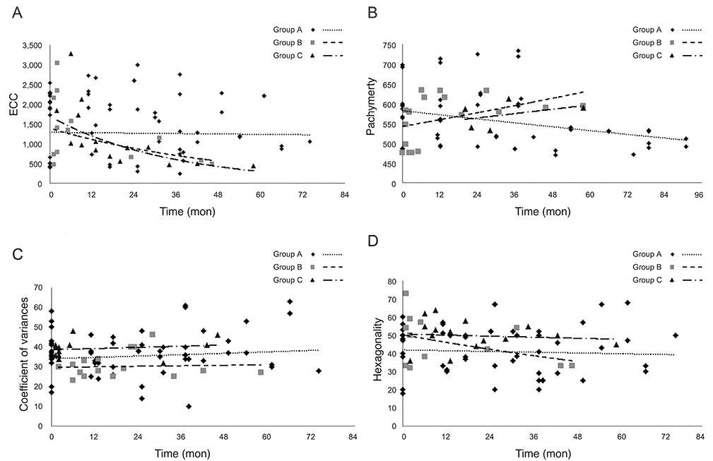
Fig. 1 Exponential curve fitting model. (A) The mean rates of endothelial cell loss were 0.82% per year, 20.39% per year, and 29.27% per year in groups A, B, and C respectively. However, only the rate in group C was statistically significant (p < 0.05). (B) The mean rates of pachymetric change were -1.81% per year, +1.21% per year, and +1.89% per year in groups A, B, and C respectively, but showed no statistical significance. (C) The mean rates of coefficient of variances change were +1.94% per year, +1.21% per year, and +0.48% per year in groups A, B, and C respectively, but showed no statistical significance. (D) The mean rates of hexagonality change were -1.04% per year, -8.06% per year, and -1.19% per year in groups A, B, and C respectively, but showed no statistical significance. ECC = endothelial cell count.
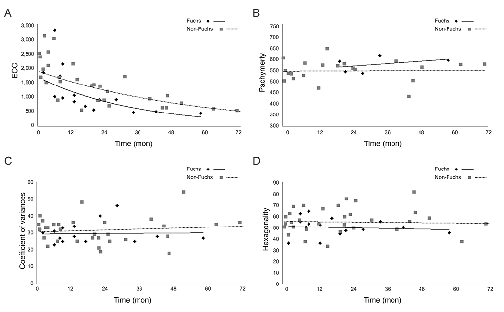
Fig. 2 Exponential curve fitting regression model in non-Fuchs patients who underwent penetrating keratoplasty and cataract surgery. (A) The mean rate of endothelial cell loss was 19.43% per year with statistical significance, while the loss rate was 29.27% per year in group C (Fuchs corneal dystrophy patients who underwent keratoplasty and cataract surgery simultaneously). (B) The mean rate of pachymetric change was +0.13% per year, and in contrast, +1.89% per year in group C. (C) The mean rate of coefficient of variances change was +1.21% per year, and +0.48% per year in group C. (D) The mean rate of hexagonality change was -0.48% per year and -1.19% per year in group C. ECC = endothelial cell count.
Reference
-
1. Borboli S, Colby K. Mechanisms of disease: Fuchs' endothelial dystrophy. Ophthalmol Clin North Am. 2002; 15:17–25.2. Adamis AP, Filatov V, Tripathi BJ, Tripathi RC. Fuchs' endothelial dystrophy of the cornea. Surv Ophthalmol. 1993; 38:149–168.3. Lietman T, Lee J, Costanza S. Those excrescences on Descemet's membrane. Br J Ophthalmol. 2003; 87:515–516.4. Cross HE, Maumenee AE, Cantolino SJ. Inheritance of Fuchs' endothelial dystrophy. Arch Ophthalmol. 1971; 85:268–272.5. Magovern M, Beauchamp GR, McTigue JW, et al. Inheritance of Fuchs' combined dystrophy. Ophthalmology. 1979; 86:1897–1923.6. Li QJ, Ashraf MF, Shen DF, et al. The role of apoptosis in the pathogenesis of Fuchs endothelial dystrophy of the cornea. Arch Ophthalmol. 2001; 119:1597–1604.7. Gottsch JD, Bowers AL, Margulies EH, et al. Serial analysis of gene expression in the corneal endothelium of Fuchs' dystrophy. Invest Ophthalmol Vis Sci. 2003; 44:594–599.8. Macnamara E, Sams GW, Smith K, et al. Aquaporin-1 expression is decreased in human and mouse corneal endothelial dysfunction. Mol Vis. 2004; 10:51–56.9. Szentmary N, Szende B, Suveges I. Epithelial cell, keratocyte, and endothelial cell apoptosis in Fuchs' dystrophy and in pseudophakic bullous keratopathy. Eur J Ophthalmol. 2005; 15:17–22.10. Bourne WM, Nelson LR, Hodge DO. Central corneal endothelial cell changes over a ten-year period. Invest Ophthalmol Vis Sci. 1997; 38:779–782.11. Roszkowska AM, Colosi P, D'Angelo P, Ferreri G. Age-related modifications of the corneal endothelium in adults. Int Ophthalmol. 2004; 25:163–166.12. Inoue K, Okugawa K, Oshika T, Amano S. Morphological study of corneal endothelium and corneal thickness in pseudoexfoliation syndrome. Jpn J Ophthalmol. 2003; 47:235–239.13. Sihota R, Lakshmaiah NC, Titiyal JS, et al. Corneal endothelial status in the subtypes of primary angle closure glaucoma. Clin Experiment Ophthalmol. 2003; 31:492–495.14. Patel SV, Hodge DO, Bourne WM. Corneal endothelium and postoperative outcomes 15 years after penetrating keratoplasty. Trans Am Ophthalmol Soc. 2004; 102:57–65.15. Ing JJ, Ing HH, Nelson LR, et al. Ten-year postoperative results of penetrating keratoplasty. Ophthalmology. 1998; 105:1855–1865.16. Seitzman GD, Gottsch JD, Stark WJ. Cataract surgery in patients with Fuchs' corneal dystrophy: expanding recommendations for cataract surgery without simultaneous keratoplasty. Ophthalmology. 2005; 112:441–446.17. Afshari NA, Pittard AB, Siddiqui A, Klintworth GK. Clinical study of Fuchs corneal endothelial dystrophy leading to penetrating keratoplasty: a 30-year experience. Arch Ophthalmol. 2006; 124:777–780.18. Seitzman GD. Cataract surgery in Fuchs' dystrophy. Curr Opin Ophthalmol. 2005; 16:241–245.19. Eghrari AO, Daoud YJ, Gottsch JD. Cataract surgery in Fuchs corneal dystrophy. Curr Opin Ophthalmol. 2010; 21:15–19.20. Traish AS, Colby KA. Approaching cataract surgery in patients with fuchs' endothelial dystrophy. Int Ophthalmol Clin. 2010; 50:1–11.
- Full Text Links
-
- Actions
-
Cited
- CITED
-
- Close
- Share
-
- Similar articles
-
- Long-Term Outcomes of Penetrating Keratoplasty in Treating Macular Corneal Dystrophy, TGFBI Dystrophy, and Fuchs' Dystrophy
- Cataract Extraction after Penetrating Keratoplasty
- Long Term Clinical Results of Penetrating Keratoplasty for Macular Corneal Dystrophy
- The Surgical Result of Phacoemulsification after Penetrating Keratoplasty
- Short-Term Outcome of Cataract Surgery Using Torsional-Mode Phacoemulsification for Patients with Low Endothelial Cell Counts
