J Korean Ophthalmol Soc.
2011 Apr;52(4):434-441.
Short-Term Outcome of Cataract Surgery Using Torsional-Mode Phacoemulsification for Patients with Low Endothelial Cell Counts
- Affiliations
-
- 1Department of Ophthalmology, Seoul National University College of Medicine, Seoul, Korea. kmk9@snu.ac.kr
- 2Seoul Artificial Eye Center, Seoul National University Hospital Clinical Research Institute, Seoul, Korea.
- 3Department of Ophthalmology, Bundang Seoul National University Hospital, Seongnam, Korea.
Abstract
- PURPOSE
To evaluate the short-term clinical outcome of cataract surgery using torsional mode phacoemulsification for patients with low endothelial cell density.
METHODS
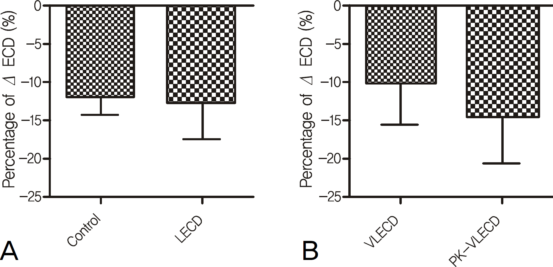
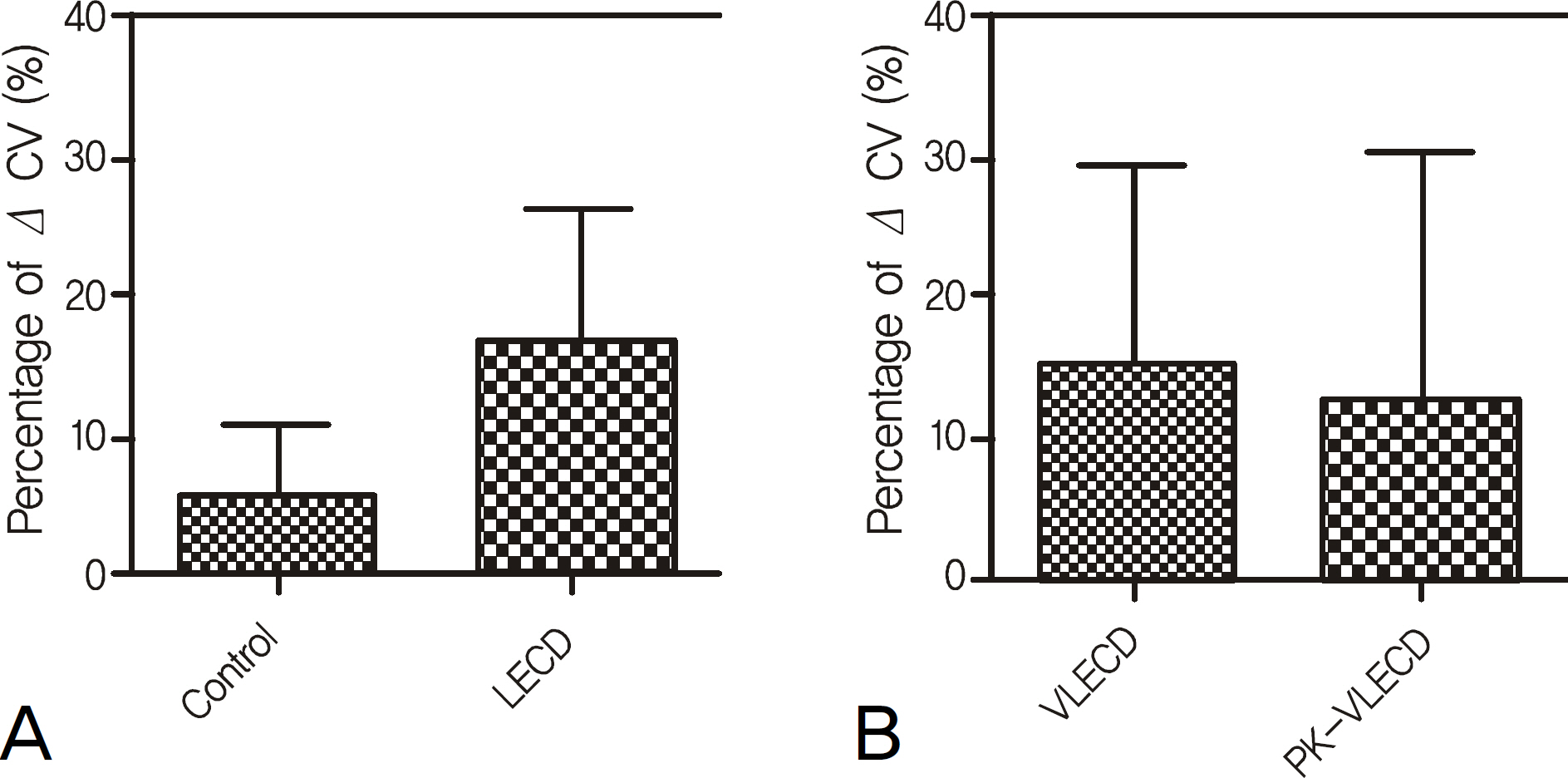
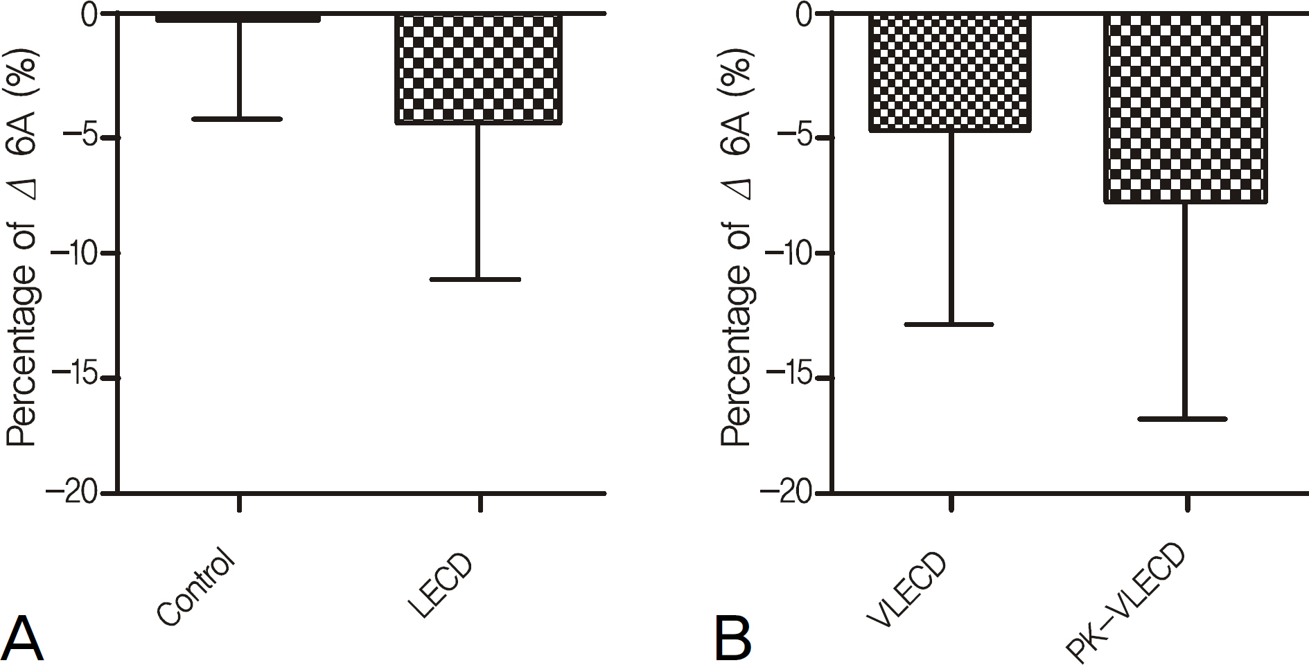
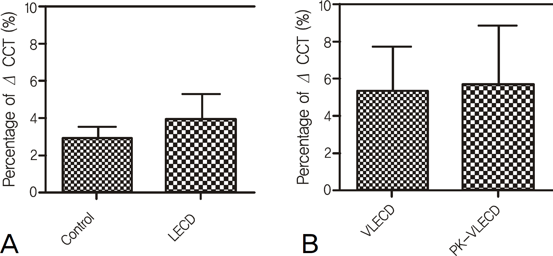
Fifty-seven eyes of 52 patients who underwent torsional phacoemulsification and intraocular lens insertion were included in the present study. Patients were divided into groups according to endothelial cell density (ECD). The control group was comprised of patients with more than 2500/mm2 of ECD and was compared with the low ECD group (LECD) comprised of patients with less than 1600/mm2 of ECD. The LECD group was further divided into a very low ECD group (VLECD) comprised of patients with less than 1000/mm2 of ECD, and a PK-VLECD group comprised of patients with less than 1000/mm2 of ECD after penetrating keratoplasty. Measurement of ECD, cell-size variation coefficient, hexagonality, and central corneal thickness were performed preoperatively and 1 month after surgery.
RESULTS
The only one patient who had undergone penetrating keratoplasty with remaining low endothelial density and grade 4 nuclear sclerosis developed overt corneal edema after cataract surgery. No statistically significant differences in the change of endothelial cell characteristics and central corneal thickness before and after surgery were observed between the control and LECD group and between the VLECD and PK-VLECD group.
CONCLUSIONS
Even in patients with low ECD, torsional phacoemulsification appears to have a similar effect on the short-term change of endothelial cells, suggesting cataract surgery can be conducted with a staged approach when the density of nucleus is moderate or less dense.
Keyword
MeSH Terms
Figure
Reference
-
References
1. Lundström M, Stenevi U, Thorburn W. The Swedish National Cataract Register: A 9-year review. Acta Ophthalmol Scand. 2002; 80:248–57.
Article2. Edwards M, Rehman S, Hood A, et al. Discharging routine phacoemulsification patients at one week. Eye. 1997; 11:850–3.
Article3. Walkow T, Anders N, Klebe S. Endothelial cell loss after phacoemulsification: relation to preoperative and intraoperative parameters. J Cataract Refract Surg. 2000; 26:727–32.
Article4. Yee RW, Matsuda M, Schultz RO, Edelhauser HF. Changes in the normal corneal endothelial cellular pattern as a function of age. Curr Eye Res. 1985; 4:671–8.
Article5. Bourne WM, Kaufman HE. Cataract extraction and the corneal endothelium. Am J Ophthalmol. 1976; 82:44–7.
Article6. Kim EC, Kim MS. A comparison of endothelial cell loss after phacoemulsification in penetrating keratoplasty patients and normal patients. Cornea. 2010; 29:510–5.
Article7. Liu Y, Zeng M, Liu X, et al. Torsional mode versus conventional ultrasound mode phacoemulsification: randomized comparative clinical study. J Cataract Refract Surg. 2007; 33:287–92.8. Kelman CD. Phacoemulsification and aspiration. A new technique of cataract removal. A preliminary report. Am J Ophthalmol. 1967; 64:23–35.9. Polack FM, Sugar A. The phacoemulsification procedure. III. Corneal complications. Invest Ophthalmol Vis Sci. 1977; 16:39–46.10. Gwin RM, Warren JK, Samuelson DA, Gum GG. Effects of phacoemulsification and extracapsular lens removal on corneal thickness and endothelial cell density in the dog. Invest Ophthalmol Vis Sci. 1983; 24:227–36.11. Sugar A, Fetherolf EC, Lin LL, et al. Endothelial cell loss from intraocular lens insertion. Ophthalmology. 1978; 85:394–9.
Article12. Kaufman HE, Katz JI. Endothelial change from intraocular lens insertion. Invest Ophthalmol Vis Sci. 1976; 15:996–1000.13. Edelhauser HF, Van Horn DL, Hyndiuk RA, Schultz RO. Intracapsular irrigating solutions, their effect on the corneal endothelium. Arch Ophthalmol. 1975; 93:648–57.14. Kim HJ, Kim JH, Lee DH. Endothelial cell damage in micro-incison cataract surgery and coaxial phacoemulsification. J Korean Ophthalmol Soc. 2007; 48:19–26.15. Alió J, Rodríguez-Prats JL, Galal A, Ramzy M. Outcomes of microincision cataract surgery versus coaxial phacoemulsification. Ophthalmology. 2005; 112:1997–2003.
Article16. Barrett G, Constable IJ. Corneal endothelial loss with new intraocular lenses. Am J Ophthalmol. 1984; 98:157–65.
Article17. El-Moatassem Kotb AM, Gamil MM. Torsional mode phacoemulsification: effective, safe cataract surgery technique of the future. Middle East Afr J Ophthalmol. 2010; 17:69–73.18. Armitage WJ, Dick AD, Bourne WM. Predicting endothelial cell loss and long-term corneal graft survival. Invest Ophthalmol Vis Sci. 2003; 44:3326–31.
Article19. Ing JJ, Ing HH, Nelson LR, et al. Ten-year postoperative results of penetrating keratoplasty. Ophthalmology. 1998; 105:1855–65.
Article20. Kim SH, Ahn BC, Chung YT. Endothelial cell changes after penetrating keratoplasty. J Korean Ophthalmol Soc. 2000; 41:1124–31.21. Wilson SE, Bourne WM, Maguire LJ, et al. Aqueous humor com-position in Fuchs’ dystrophy. Invest Ophthalmol Vis Sci. 1989; 30:449–53.22. Giasson CJ, Solomon LD, Polse KA. Morphometry of corneal endothelium in patients with corneal guttata. Ophthalmology. 2007; 114:1469–75.
Article23. Sugar J, Mitchelson J, Kraff M. The effect of phacoemulsification on corneal endothelial cell density. Arch Ophthalmol. 1978; 96:446–8.
Article24. Matsuda M, Miyake K, Inaba M. Long-term corneal endothelial changes after intraocular lens implantation. Am J Ophthalmol. 1988; 105:248–52.
Article25. Schultz RO, Glasser DB, Matsuda M, et al. Response of the corneal endothelium to cataract surgery. Arch Ophthalmol. 1986; 104:1164–9.
Article
- Full Text Links
- Actions
-
Cited
- CITED
-
- Close
- Share
- Similar articles
-
- Comparison of Clinical Results Between OZil(R) Mode and Hyperpulse Mode in Phacoemulsification
- Comparison of Clinical Outcomes Between Torsional Phacoemulsification of Infiniti(R) and Longitudinal Phacoemulification of Stellaris(R) Through 2.2 mm Microincision
- The Comparison between Torsional and Conventional Mode Phacoemulsification in Moderate and Hard Cataracts
- Clinical Outcomes of Cataract Surgery Using Torsional Mode Phacoemulsification and Soft Shell Technique
- Comparison of Surgical Results between WhiteStar Mode and Continuous Mode in the Phacoemulsification Unit


