Decreasing Incidence of Trichophyton mentagrophytes in Korea: Analysis of 6,250 Cases during the Last 21-Year-Period (1992-2012)
- Affiliations
-
- 1Department of Dermatology, Kyungpook National University School of Medicine, Daegu, Korea. weonju@knu.ac.kr
- 2Institute of Medical Mycology, Catholic Skin Clinic, Daegu, Korea.
- KMID: 1789987
- DOI: http://doi.org/10.3346/jkms.2014.29.2.272
Abstract
- Trichophyton mentagrophytes is the second common dermatophyte in Korea. However, few reports have been issued on the epidemiological and mycological characteristics of T. mentagrophytes in Korea based on long-term, large-scale study. The purpose of this study was to elucidate the epidemiological and mycological characteristics of T. mentagrophytes in Korea. During the 21-yr-period from 1992 to 2012, 6,250 patients with T. mentagrophytes were surveyed to determine annual incidence and the distribution of subjects by age, sex, season, involved sites, and place of residence. T. mentagrophytes infections were confirmed by fungal culture. In addition, the colony appearance of T. mentagrophytes was classified as granular, persicolor, powdery, or downy. Epidemiological analysis showed that annual incidence reached a peak in 2005, and then gradually decreased. T. mentagrophytes infection was most common in July, and was found predominantly in middle-aged adults, especially in those in their forties. Mycological analysis showed a powdery colony appearance was the most common, followed by persicolor and granular colonies. Toewebs were most frequently involved. This investigation on T. mentagrophytes provides insights into its incidence and characteristics.
Keyword
MeSH Terms
Figure
-

Fig. 1 Culture appearances of T. mentagrophytes colonies. (A) Persicolor. (B) Downy. (C) Powdery. (D) Granular.

Fig. 2 Annual incidences of dermatophytoses caused by T. mentagrophytes from 1992 to 2012.

Fig. 3 Distribution of patients with dermatophytosis caused by T. mentagrophytes by age and sex from 1992 to 2012.
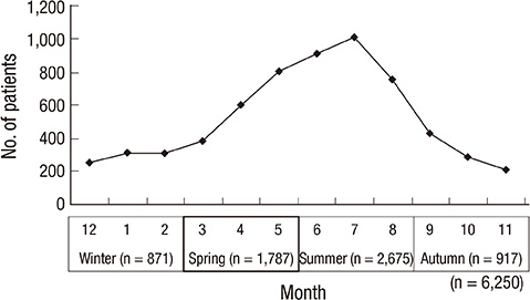
Fig. 4 Monthly and seasonal incidences of dermatophytoses caused by T. mentagrophytes from 1992 to 2012.

Fig. 5 Clinical appearance of the interdigital subtype of tinea pedis. (A) Scales on toewebs and of the vesicular subtype, (B) Grouped vesicles on a sole.
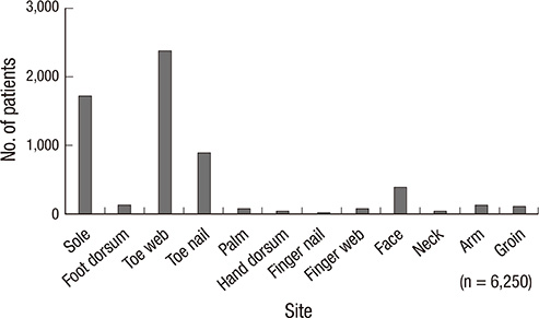
Fig. 6 Distribution of patients with dermatophytosis caused by T. mentagrophytes by involved site.
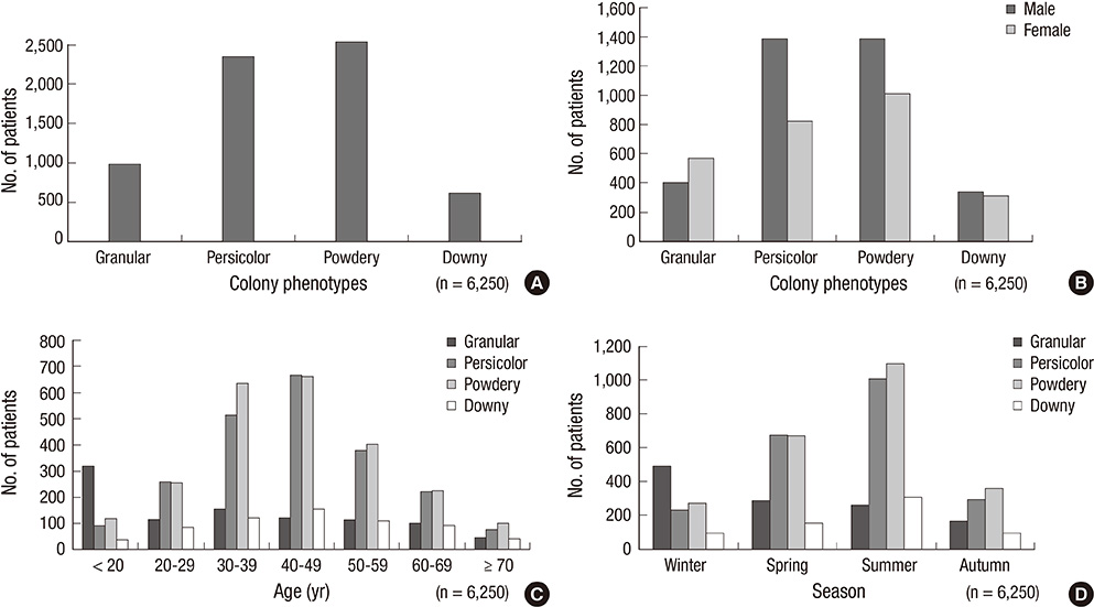
Fig. 7 Frequency of T. mentagrophytes by colony phenotype (A). Distribution of colony phenotypes of T. mentagrophytes by sex (B), age (C), and season (D).
Cited by 2 articles
-
The Epidemiology of Dermatophyte Infection in Southeastern Korea (1979~2013)
Sang Lim Kim, Kyou Chae Lee, Yong Hyun Jang, Seok-Jong Lee, Do Won Kim, Weon Ju Lee, Yong Jun Bang, Jae Bok Jun
Ann Dermatol. 2016;28(4):524-527. doi: 10.5021/ad.2016.28.4.524.Low But Continuous Occurrence of
Microsporum gypseum Infection in the Study on 198 Cases in South Korea from 1979 to 2016
Weon Ju Lee, Jun Hong Park, Jun Young Kim, Yong Hyun Jang, Seok-Jong Lee, Yong Jun Bang, Jae Bok Jun
Ann Dermatol. 2018;30(4):427-431. doi: 10.5021/ad.2018.30.4.427.
Reference
-
1. Kim BS, Suh SB. Mycological and clinical observations on dermatophytosis. Korean J Dermatol. 1976; 14:325–334.2. Moon HJ, Lee JB, Kim SJ, Lee SC, Won YH. Clinical and mycological studies on dermatomycosis (1991-2000). Korean J Med Mycol. 2002; 7:78–85.3. Kim KH. Dermatophytes isolated from Korea. Yeungnam Univ J Med. 1990; 7:13–26.4. Rippon JW. Medical Mycology: the pathogenic fungi and the pathogenic actinomycetes. 3rd ed. Philadelphia: WB Saunders;1988. p. 121–275.5. Emmons CW. Dermatophytes: natural groupings based on the form of the spores and accessory organs. Arch Derm Syphilol. 1934; 30:337–362.6. Schieke SM, Garg A. Superficial fungal infection. In : Goldsmith LA, Kats SI, Gichrest BA, Paller AS, Leffell DJ, Wolff K, editors. Fitzpatrick's dermatology in general medicine. 8th ed. New York: McGraw-Hill;2012. p. 2277–2297.7. Choe YS, Park BC, Lee WJ, Jun JB, Suh SB, Bang YJ. The clinical observation of Trichophyton verrucosum infections during the last 19 years (1986-2004). Korean J Med Mycol. 2006; 11:45–53.8. Lee WJ, Song CH, Lee SJ, Kim DW, Jun JB, Bang YJ. Decreasing prevalence of Microsporum canis infection in Korea: through analysis of 944 cases (1993-2009) and review of our previous data (1975-1992). Mycopathologia. 2012; 173:235–239.9. Suh SB, Kim SW, Oh SH, Choi SK, Bang YJ. A case of black dot ringworm caused by Trichophyton tonsurans. Korean J Dermatol. 1998; 36:918–923.10. Park JS, Kim SW, Jun JB, Suh SB, Bang YJ. Clinical and epidemiologic study of Trichophyton tonsurans infections (1995-2003). Korean J Med Mycol. 2004; 9:197–205.11. Kim KH, Moon BC, Choi JS. Subtypes and mycologic characteristics of Trichopyhton genus isolated in Taegu Korea. Korean J Med Mycol. 1997; 2:129–143.12. Kim KS, Shin DH, Bang YJ, Choi JS, Kim KH. Mycologic findings of Trichophyton mentagrophytes var. mentagrophytes isolated from the patients with dermatophytosis in Taegu area and microsporum persicolor. Korean J Med Mycol. 1999; 4:109–116.13. St-Germain G, Summerbell R. Identifying filamentous fungi: a clinical laboratory handbook. Belmont: Star Publishing;1996. p. 212–213.14. Summerbell RC, Kane J. The genera Trichophyton and Epidermophyton. In : Kane J, Summerbell RC, Sigler L, Krajden S, Land G, editors. Laboratory handbook of dermatophytes. Belmont: Star Publishing Co;1997. p. 131–192.15. Singh D, Patel DC, Rogers K, Wood N, Riley D, Morris AJ. Epidemiology of dermatophyte infection in Auckland, New Zealand. Australas J Dermatol. 2003; 44:263–266.16. Morishita N, Ninomiya J, Sei Y, Takiuchi I. Effects of temperature, humidity, minor injury and washing on penetration of dermatophytes into human stratum corneum. Nihon Ishinkin Gakkai Zasshi. 2003; 44:269–271.
- Full Text Links
-
- Actions
-
Cited
- CITED
-
- Close
- Share
-
- Similar articles
-
- Tinea Manuum Caused by Trichophyton mentagrophytes var. erinacei
- Two Cases of Fungal Granuloma Caused by Trichophyton mentagrophytes
- Mycologic Findings of Trichophyton mentagrophytes var. mentagrophytes Isolated from the Patients with Dermatophytosis in Taegu Area and Microsporum Persicolor
- A Case of Kerion Celsi Caused by Trichophyton mentagrophytes
- Plate Medium Findings of Trichophyton mentagrophytes
