Yonsei Med J.
2007 Dec;48(6):1066-1071. 10.3349/ymj.2007.48.6.1066.
Extrapulmonary Small Cell Carcinoma of the Liver: Clinicopathological and Immunohistochemical Findings
- Affiliations
-
- 1Department of Pathology, Inha University College of Medicine, Incheon, Korea. jmkpath@inha.ac.kr
- 2Department of General Surgery, Inha University College of Medicine, Incheon, Korea.
- 3Department of Hematooncology, Inha University College of Medicine, Incheon, Korea.
- KMID: 1786228
- DOI: http://doi.org/10.3349/ymj.2007.48.6.1066
Abstract
- Patients with primary small cell carcinoma of the liver have rarely been described in medical literature. Knowledge of clinical, pathological and immunohistochemical properties remains limited. We described an 82-year-old female patient with primary small cell carcinoma of the liver. Histologically, the tumor showed typical morphology of a pulmonary small cell carcinoma. Immunohistochemically, the tumor revealed neuroendocrine differentiation; positive reaction for chromogranin, synaptophysin, CD56, and neuron specific enolase. The tumor was also positive for TTF-1 and c-kit but completely negative for hepatocyte, carcinoembryonic antigen, cytokeratin 7; 19; and 20. Herein, we discussed the clinical, pathological and immunohistochemical findings of extrapulmonary small cell carcinoma of the liver and reviewed the relevant literature.
MeSH Terms
Figure
-
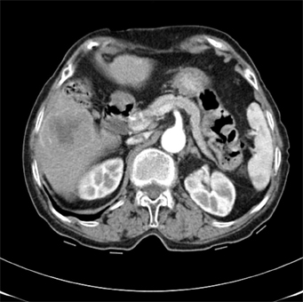
Fig. 1 Abdominal CT scan shows a 5.6 cm-size liver mass with peripheral rim enhancement.
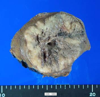
Fig. 2 Grossly, the well demarcated tumor shows central necrosis and cystic change.

Fig. 3 Microscopic findings of the tumor reveal solid small round cells and necrosis (A). The tumor cells show oval to fusiform hyperchromatic nuclei and indistinct nucleoli with frequent mitoses (B).

Fig. 4 Immunohistochemical staining for CD56 (A) and synaptophysin (B) reveals a strong positive reaction.
Reference
-
1. Richardson RL, Weiland LH. Undifferentiated small cell carcinomas in extrapulmonary sites. Semin Oncol. 1982. 8:484–496.2. Ryu SH, Han SY, Suh SH, Koo YH, Cho JH, Han SH, et al. A case of primary small cell carcinoma of the liver. Korean J Hepatol. 2005. 11:289–292.3. Kim YH, Kwon R, Jung GJ, Roh MH, Han SY, Kwon HC, et al. Extrapulmonary small-cell carcinoma of the liver. J Hepatobiliary Pancreat Surg. 2004. 11:333–337.4. Sengoz M, Abacioglu U, Salepci T, Eren F, Yumuk F, Turhal S. Extrapulmonary small cell carcinoma: multimodality treatment results. Tumori. 2003. 89:274–277.5. Zanconati F, Falconieri G, Lamovec J, Zidar A. Small cell carcinoma of the liver: a hitherto unreported variant of hepatocellular carcinoma. Histopathology. 1996. 29:449–453.6. Kim KJ, Yim HJ, Kim MJ, Choung RS, Yeon JE, Lee HS, et al. A case of primary small cell neuroendocrine carcinoma of the liver. Korean J Gastroenterol. 2006. 48:37–41.7. Kim KO, Lee HY, Chun SH, Shin SJ, Kim MK, Lee KH, et al. Clinical overview of extrapulmonary small cell carcinoma. J Korean Med Sci. 2006. 21:833–837.8. Vrouvas J, Ash DV. Extrapulmonary small cell cancer. Clin Oncol (R Coll Radiol). 1995. 7:377–381.9. Dala R, Shoosmith J, Lilenbaum R, Cabello-Inchausti B. Primary hepatic neuroendocrine carcinoma: an underdiagnosed entity. Ann Diagn Pathol. 2006. 10:28–31.10. Hsueh C, Tan XD, Gonzalez-Crussi F. Primary hepatic neuroendocrine carcinoma in a child. Morphologic, immunocytochemical, and molecular biologic studies. Cancer. 1993. 71:2660–2665.11. Pilichowska M, Kimura N, Ouchi A, Lin H, Mizuno Y, Nagura H. Primary hepatic carcinoid and neuroendocrine carcinoma: clinicopathological and immunohistochemical study of five cases. Pathol Int. 1999. 49:318–324.12. Kaya G, Pasche C, Osterheld MC, Chaubert P, Fontolliet C. Primary neuroendocrine carcinoma of the liver: an autopsy case. Pathol Int. 2001. 51:874–878.13. Brenner B, Tang LH, Klimstra DS, Kelsen DP. Small-cell carcinomas of the gastrointestinal tract: a review. J Clin Oncol. 2004. 22:2730–2739.14. Ordonez NG. Value of thyroid transcription factor-1 immunostaining in distinguishing small cell lung carcinomas from other small cell carcinomas. Am J Surg Pathol. 2000. 24:1217–1223.15. Kaufmann O, Dietel M. Expression of thyroid transcription factor-1 in pulmonary and extrapulmonary small cell carcinomas and other neuroendocrine carcinomas of various primary sites. Histopathology. 2000. 36:415–420.16. Jones TD, Kernek KM, Yang XJ, Lopez-Beltran A, MacLennan GT, Eble JN, et al. Thyroid transcription factor 1 expression in small cell carcinoma of the urinary bladder: an immunohistochemical profile of 44 cases. Hum Pathol. 2005. 36:718–723.17. Yao JL, Madeb R, Bourne P, Lei J, Yang X, Tickoo S, et al. Small cell carcinoma of the prostate: an immunohistochemical study. Am J Surg Pathol. 2006. 30:705–712.
- Full Text Links
-
- Actions
-
Cited
- CITED
-
- Close
- Share
-
- Similar articles
-
- A Case of Small Cell Carcinoma of the Pleura
- A case of primary small cell neuroendocrine carcinoma of the liver
- A Case of Small Cell Neuroendocrine Carcinoma in the Parotid and Lacrimal Glands
- Primary small cell carcinoma of the esophagus and stomach: report of two cases and review of extrapulmonary small cell carcinoma in Korea
- A case of primary small-cell carcinoma of the larynx
