The Correlation between F-wave Motor Unit Number Estimation (F-MUNE) and Functional Recovery in Stroke Patients
- Affiliations
-
- 1Department of Physical & Rehabilitation Medicine, Research Institute of Medical Sciences, Chonnam National University Medical School & Hospital, Gwangju, Korea. sam91@jnu.ac.kr
- KMID: 1785761
- DOI: http://doi.org/10.3346/jkms.2007.22.6.1002
Abstract
- The aim of this study was to follow up the changes in the number of motor units according to the Brunnstrom stage through a motor unit number estimation of the Fwave (F-MUNE) after a stroke, and to identify the functional significance of F-MUNE. Twenty-five patients (15 men, 10 women) with a first unilateral stroke were recruited. The maximal M-potential was evoked by the supramaximal stimulation of the median nerve at the wrist, and the maximal stimulation intensity was determined on both hemiplegic and unaffected hands. The reproducible all-or-none F-wave was evoked in 30% of the maximal stimulation intensity and was constantly stimulated at that level. The prototypes of the F-wave were chosen, and the values of F-MUNE were calculated by dividing the amplitude of the maximal M-potential by the mean amplitude of the F-prototype. The changes in F-MUNE were compared according to the progression of the Brunnstrom stage and correlated with those of the functional scales. The mean motor unit numbers decreased significantly in the hemiplegic side compared with the unaffected side. According to the progression of the Brunnstrom stage, the values of F-MUNE were reduced significantly by increasing the amplitude and recruitment of the F-prototype, and the functional scores also improved. These results show that the F-MUNE equation did not show a functional recoveryrelated increase in stroke patients.
MeSH Terms
Figure
-
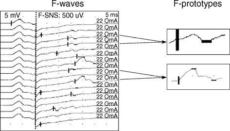
Fig. 1 The prototypes of the F-wave, identically matched two or more F-waves with the same latency, amplitude, and shape were selected among the entire products of individual F-waves.
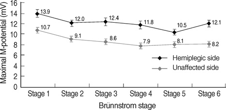
Fig. 2 The peak amplitude of the maximal M-potential (MMP) in the hemiplegic side was significantly smaller than in the unaffected side (p=0.000, between groups), but there was no significant changes according to the progression of the Brünnstrom stage on both sides (p>0.05, within groups). Repetitive measure analysis was used to examine the significance of the serial changes in the MMP according to the progression of the Brünnstrom stage.

Fig. 3 The functional scores by the functional independence measure (FIM) and modified Barthel index (MBI) were significantly improved according to the progression of the Brünnstrom stage (p= 0.000, repetitive measure analysis).
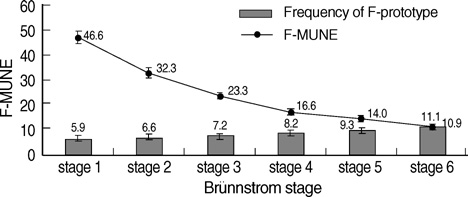
Fig. 4 The frequency of the F-prototypes was increased progressively and estimation of the number of motor units showing the F-wave (F-MUNE) decreased significantly according to the progression of the Brünnstrom stage (p=0.000, repetitive measure analysis).
Cited by 1 articles
-
Effect of Motor Imagery on the F-Wave Parameters in Hemiparetic Stroke Survivors
Mahshid Naseri, Peyman Petramfar, Alireza Ashraf
Ann Rehabil Med. 2015;39(3):401-408. doi: 10.5535/arm.2015.39.3.401.
Reference
-
1. Jorgensen HS, Nakayama H, Raaschou HO, Vive-Larsen J, Støier M, Olsen TS. Outcome and time course of recovery in stroke. Part 1: Outcome. The Copenhagen Stroke Study. Arch Phys Med Rehabil. 1995. 76:399–405. .2. Fisher MA, Shahani BT, Young RR. Assessing segmental excitability after acute rostral lesions: II. The blink reflex. Neurology. 1979. 29:45–50.3. Drory VE, Neufeld MY, Korczyn AD. F wave characteristics following acute and chronic upper motor neuron lesions. Electromyogr Clin Neurophysiol. 1993. 33:441–446.4. McComas AJ, Sica REP, Upton AR, Aguilera N, Currie S. Motoneuron dysfunction in patients with hemiplegic atrophy. Nat New Biol. 1971. 233:21–23.5. Higashi T, Funase K, Kusano K, Tabira T, Harada N, Sakakibara A, Yashimura T. Motoneuron pool excitability of hemiplegic patients: assessing recovery stages by using H-reflex and M response. Arch Phys Med Rehabil. 2001. 82:1604–1610.
Article6. Burke D. Critical examination of the case for or against fusimotor involvement in disorders of muscle tone. Adv Neurol. 1983. 39:133–150.7. Sohn MK, Han SM. Facilitation of nerve conduction by distant muscle contraction in stroke patients. J Korean Acad Rehabil Med. 2005. 29:50–57.8. McComas AJ. Invited review. Motor unit estimation: methods, results, and present status. Muscle Nerve. 1991. 14:585–597.
Article9. Stashuk DW, Doherty TJ, Kassam A, Brown WF. Motor unit number estimates based on the automated analysis of F-responses. Muscle Nerve. 1994. 17:881–890.
Article10. Hara Y, Akaboshi K, Masakado Y, Chino N. Physiological decrease of single thenar motor units in the F-response in stroke patients. Arch Phys Med Rehabil. 2000. 81:418–423.11. Doherty TJ, Brown WF. The estimated numbers and relative sizes of thenar motor units as selected by multiple point stimulation in young and older adults. Muscle Nerve. 1993. 16:355–366.
Article12. Benecke R, Berthold A, Conrad B. Denervation activity in the EMG of patients with upper motor neuron lesions: time course, local distribution and pathogenetic aspects. J Neurol. 1983. 230:143–151.
Article13. Dattola R, Girlanda P, Vita G, Snatoro M, Robert ML, Toscano A, Venuto C, Baradello A, Messina C. Muscle rearrangement in patients with hemiparesis after stroke: an electrophysiological and morphological study. Eur Neurol. 1993. 33:109–114.
Article14. Brunnstrom S. Motor testing procedures in hemiplegia: Based on sequential recovery stages. Phys Ther. 1966. 46:357–375.
Article15. Shah S, Vanclay F, Cooper B. Improving the sensitivity of the Barthel Index for stroke rehabilitation. J Clin Epidemiol. 1989. 42:703–709.
Article16. Magladery JW, McDougal DB. Electrophysiological studies of nerve and reflex activity in normal man. I. Identification of certain reflexes in the electromyogram and the conduction velocity of peripheral nerve fibers. Bull Johns Hopkins Hosp. 1950. 86:265–290.17. Fisher MA. Inhibition of motoneuron discharge by peripheral nerve stimulation: an F-response analysis. Muscle Nerve. 1991. 14:120–123.18. Kimura J. . Other techniques to assess nerve function, Electrodiagnosis in diseases of nerve and muscle: principles and practice. 1983. 3rd ed. Philadelphia: F.A. Davis;216–217.19. Manganotti P, Patuzzo S, Cortese F, Palermo A, Smania N, Fiaschi A. Motor disinhibition in affected and unaffected hemisphere in the early period of recovery after stroke. Clin Neurophysiol. 2002. 113:936–943.
Article20. Kondo A, Nagara H, Tateishi J. A morphometric study of myelinated fibers in the fifth lumbar ventral roots in patients with cerebrovascular diseases. Clin Neuropathol. 1987. 6:250–256.21. Spaans F, Wilts G. Denervation due to lesions of the central nervous system. An EMG study in cases of cerebral contusion and cerebrovascular accidents. J Neurol Sci. 1982. 57:291–305.22. Fierro B, Raimondo D, Modica A. Analysis of F response in upper motoneuron lesions. Acta Neurol Scand. 1990. 82:329–334.23. Yan K, Fang J, Turan b, Chira-Adisai W, Shahani BT. The use of fractional parameter in analyzing motor unit discharge pattern in stroke patients: a correlation with the functional independence measurement. Electromyogr Clin Neurophysiol. 2000. 40:3–9.
- Full Text Links
-
- Actions
-
Cited
- CITED
-
- Close
- Share
-
- Similar articles
-
- Motor Unit Number Estimation in Thenar Muscles of the Hemiplegic Patients
- Motor Unit Number Estimation of the Thenar Muscles
- Estimation of Motor Unit Number According to Severity of Peripheral Nerve Injury in Rat
- Motor Unit Numbers Estimation in Abductor Pollicis Brevis Muscle of Normal Adult
- Motor Unit Number Estimation in Evaluating Disease Progression in Patients with Amyotrophic Lateral Sclerosis
