Optimization of MR Parameters of 3D TOF-MRA for Various Intracranial Stents at 3.0T MRI
- Affiliations
-
- 1Department of Radiology, Konkuk University Medical Center, Konkuk University School of Medicine, Seoul, Korea. hgroh@kuh.ac.kr
- 2Department of Neurosurgery, Konkuk University Medical Center, Konkuk University School of Medicine, Seoul, Korea.
- 3Department of Radiolgy, Konkuk University Medical Center, Seoul, Korea.
- KMID: 1783986
- DOI: http://doi.org/10.5469/neuroint.2011.6.2.71
Abstract
- PURPOSE
The in-stent signal reduction of the stented artery caused by susceptibility artifact or radiofrequency shielding artifact limited the use of time-of-flight MR angiography (TOF-MRA) as a follow-up tool after intracranial stenting. We showed the degree of an artifact according to different stent types, and optimized MR parameters for TOF-MRA in patients with intracranial stent on 3.0 T MRI.
MATERIALS AND METHODS
Four stents (Neuroform, Wingspan, Solitaire, and Enterprise) were placed in a vascular flow phantom and imaged by changing flip angle (FA; 20degrees,30degrees,40degrees,50degrees and 60degrees) and bandwidth (BW; 31, 42 and 62.5 KHz) using TOF-MRA. Source data of each image set with different FA and BW were reconstructed with the maximal intensity projection (MIP) technique, and MIP images were used to evaluate the in-stent signal reduction of each stent according to the change of MR parameters. The in-stent signal reduction was assessed by calculating the relative in-stent signal (RIS) inside the stent as compared with background and signal intensity of the tube outside the stent. The optimal FA and BW of each stent were determined by comparing the RIS in each stent by one-sample t test. Finally, one neuroradiologist chose one image set with the best image quality.
RESULTS
The mean RIS for Neuroform, Wingspan, Solitaire and Enterprise stent was 66.3 +/- 6.0, 44.2 +/- 5.8, 22.8 +/- 3.3 and 8.2 +/- 2.9, respectively. The significantly high RIS of each stent was obtained with FA/BW value of 20degrees/31 KHz (Neuroform), 20degrees/31 KHz and 30degrees/42 KHz (Wingspan), 40degrees/42 KHz and 50degrees/31 KHz (Solitaire) and 40degrees/31 KHz and 50degrees/31 KHz (Enterprise). Among these MIP images with significantly high RIS, images with FA/BW value of 20degrees/31 KHz (Neuroform and Wingspan) and 50degrees/31 KHz (Solitaire and Enterprise) had the best image quality.
CONCLUSION
The degree of artifact was variable according to the design of each intracranial stent. The luminal visualization of closed-cell design stents such as Solitaire and Enterprise can be improved by higher FA. Thus, MR parameter should be adjusted according to the type of intracranial stents.
MeSH Terms
Figure
-
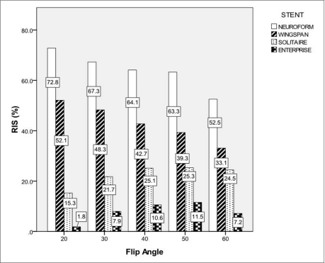
Fig. 1 Relative In-stent Signal According to the Change of FA.Bars represent RIS for each stent according to the change of FA at BW of 31 KHz. The number in the boxes means RIS value for each stent.

Fig. 2 Mean Relative In-stent Signal According to the Change of Bandwidth.Bars represent mean RIS (mean of RIS at FA of 30° and 40°) for each stent according to the change of BW. The number in the boxes means mean RIS value for each stent.
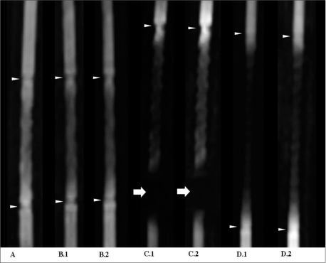
Fig. 3 MIP images of the each stent with significantly high RIS. MIP images of Neuroform (A, FA/BW; 20°/31 KHz), Wingspan (B.1, FA/BW; 20°/31 KHz, and B.2, FA/BW; 30°/42 KHz), Solitaire (C.1, FA/BW; 40°/42 KHz, and C.2, FA/BW; 50°/31 KHz) and Enterprise (D.1, FA/BW; 40°/31 KHz, and D.2, FA/BW; 50°/31 KHz). A band-like artifact (arrowhead) is visible at the end of the each stent due to platinum marker. A complete signal loss of the lumen (arrow) near the proximal marker of the Solitaire stent was shown.
Cited by 1 articles
-
Time-of-Flight Magnetic Resonance Angiography for Follow-Up of Coil Embolization with Enterprise Stent for Intracranial Aneurysm: Usefulness of Source Images
Young Dae Cho, Kang Min Kim, Woong Jae Lee, Chul-Ho Sohn, Hyun-Seung Kang, Jeong Eun Kim, Moon Hee Han
Korean J Radiol. 2014;15(1):161-168. doi: 10.3348/kjr.2014.15.1.161.
Reference
-
1. Tähtinen OI, Vanninen RL, Manninen HI, Rautio R, Haapanen A, Niskakangas T, et al. Wide-necked intracranial aneurysms: Treatment with stent-assisted coil embolization during acute (<72 hours) subarachnoid hemorrhage-experience in 61 consecutive patients1. Radiology. 2009; 253:199–208. PMID: 19710006.2. Fiorella D, Albuquerque FC, Deshmukh VR, McDougall CG. Usefulness of the neuroform stent for the treatment of cerebral aneurysms: results at initial (3-6-mo) follow-up. Neurosurgery. 2005; 56:1191–1202. PMID: 15918935.
Article3. Klisch J, Eger C, Sychra V, Strasilla C, Basche S, Weber J. Stent-assisted coil embolization of posterior circulation aneurysms using solitaire ab: preliminary experience. Neurosurgery. 2009; 65:258–266. discussion 266. PMID: 19625903.4. Lubicz B, Collignon L, Raphaeli G, Bandeira A, Bruneau M, De Witte O. Solitaire stent for endovascular treatment of intracranial aneurysms: immediate and mid-term results in 15 patients with 17 aneurysms. J Neuroradiol. 2010; 37:83–88. PMID: 20381147.
Article5. Suh DC, Kim JK, Choi JW, Choi BS, Pyun HW, Choi YJ, et al. Intracranial stenting of severe symptomatic intracranial stenosis: results of 100 consecutive patients. AJNR Am J Neuroradiol. 2008; 29:781–785. PMID: 18310234.
Article6. Costalat V, Maldonado IL, Vendrell JF, Riquelme C, Machi P, Arteaga C, et al. Endovascular treatment of symptomatic intracranial stenosis with the wingspan stent system and gateway pta balloon: a multicenter series of 60 patients with acute and midterm results. J Neurosurg. 2011.
Article7. Jabbour PM, Tjoumakaris SI, Rosenwasser RH. Endovascular management of intracranial aneurysms. Neurosurg Clin N Am. 2009; 20:383–398. PMID: 19853799.
Article8. Hwang J, Roh H, Chun Y, Kang HS, Choi J, Moon WJ, et al. Endovascular coil embolization of very small intracranial aneurysms. Neuroradiology. 2011; 53:349–357. PMID: 20574735.
Article9. Klisch J, Clajus C, Sychra V, Eger C, Strasilla C, Rosahl S, et al. Coil embolization of anterior circulation aneurysms supported by the solitaire™, ab neurovascular remodeling device. Neuroradiology. 2010; 52:349–359. PMID: 19644683.
Article10. Kaufmann TJ, Huston J, Mandrekar JN, Schleck CD, Thielen KR, Kallmes DF. Complications of diagnostic cerebral angiography: evaluation of 19?826 consecutive patients1. Radiology. 2007; 243:812–819. PMID: 17517935.
Article11. Lakshminarayan R, Simpson J, Ettles D. Magnetic resonance angiography: current status in the planning and follow-up of endovascular treatment in lower-limb arterial disease. Cardiovasc Intervent Radiol. 2009; 32:397–405. PMID: 19130124.
Article12. Hamer OW, Finkenzeller T, Borisch I, Paetzel C, Zorger N, Feuerbach S, et al. In vivo evaluation of patency and in-stent stenoses after implantation of nitinol stents in iliac arteries using MR angiography. AJR Am J Roentgenol. 2005; 185:1282–1288. PMID: 16247150.
Article13. Lettau M, Sauer A, Heiland S, Rohde S, Bendszus M, Hahnel S. Carotid artery stents: in vitro comparison of different stent designs and sizes using CT angiography and contrast-enhanced MR angiography at 1.5t and 3t. AJNR Am J Neuroradiol. 2009; 30:1993–1997. PMID: 19749216.
Article14. Girardi M, Kay J, Elston DM, Leboit PE, Abu-Alfa A, Cowper SE. Nephrogenic systemic fibrosis: clinicopathological definition and workup recommendations. J Am Acad Dermatol. 2011; 7. 01. [Epub ahead of print].
Article15. Yamada N, Hayashi K, Murao K, Higashi M, Iihara K. Time-of-flight MR angiography targeted to coiled intracranial aneurysms is more sensitive to residual flow than is digital subtraction angiography. AJNR Am J Neuroradiol. 2004; 25:1154–1157. PMID: 15313700.16. Wallace RC, Karis JP, Partovi S, Fiorella D. Noninvasive imaging of treated cerebral aneurysms, part i: MR angiographic follow-up of coiled aneurysms. AJNR Am J Neuroradiol. 2007; 28:1001–1008. PMID: 17569946.
Article17. Trost DW, Zhang HL, Prince MR, Winchester PA, Wang Y, Watts R, et al. Three-dimensional MR angiography in imaging platinum alloy stents. J Magn Reson Imaging. 2004; 20:975–980. PMID: 15558574.
Article18. Lenhart M, Völk M, Manke C, Nitz WR, Strotzer M, Feuerbach S, et al. Stent appearance at contrast-enhanced mr angiography: in vitro examination with 14 stents. Radiology. 2000; 217:173–178. PMID: 11012441.
Article19. Wang Y, Truong TN, Yen C, Bilecen D, Watts R, Trost DW, et al. Quantitative evaluation of susceptibility and shielding effects of nitinol, platinum, cobalt-alloy, and stainless steel stents. Magn Reson Med. 2003; 49:972–976. PMID: 12704782.
Article20. Lovblad KO, Yilmaz H, Chouiter A, San Millan Ruiz D, Abdo G, Bijlenga P, et al. Intracranial aneurysm stenting: follow-up with MR angiography. J Magn Reson Imaging. 2006; 24:418–422. PMID: 16795090.21. Bartels LW, Bakker CJG, Viergever MA. Improved lumen visualization in metallic vascular implants by reducing rf artifacts. Magnetic Resonance in Medicine. 2002; 47:171–180. PMID: 11754456.
Article22. van Holten J, Wielopolski P, Bruck E, Pattynama PM, van Dijk LC. High flip angle imaging of metallic stents: implications for MR angiography and intraluminal signal interpretation. Magn Reson Med. 2003; 50:879–883. PMID: 14523976.
Article23. Frölich A, Pilgram-Pastor S, Psychogios M, Mohr A, Knauth M. Comparing different MR angiography strategies of carotid stents in a vascular flow model: toward stent-specific recommendations in MR follow-up. Neuroradiology. 2011; 53:359–365. PMID: 20721544.
Article24. Wall A, Kugel H, Bachman R, Matuszewski L, Krämer S, Heindel W, et al. 3.0 t vs. 1.5 t MR angiography: in vitro comparison of intravascular stent artifacts. J Magn Reson Imaging. 2005; 22:772–779. PMID: 16270296.
Article
- Full Text Links
-
- Actions
-
Cited
- CITED
-
- Close
- Share
-
- Similar articles
-
- Time-Resolved 3D Contrast-Enhanced MRA on 3.0T: a Non-Invasive Follow-Up Technique after Stent-Assisted Coil Embolization of the Intracranial Aneurysm
- Usefulness of MR Angiography in Patients with Non-Traumatic Intracranial Hemorrhagic DiseasesI
- Comparison of 3D TOF MRA with Contrast Enhanced MRA in Intracranial Atherosclerotic Occlusive Disease
- Usefulness of three-dimensional fast imaging employing steady-state acquisition MRI of large vessel occlusion for detecting occluded middle cerebral artery and internal carotid artery before acute mechanical thrombectomy
- Image Quality of the 3 Dimensional Phase-Contrast Technique in an Intracranial Magnetic Resonance Angiography with Artifacts Caused by Orthodontic Devices: A Comparison with 3 Dimensional Time-of-Flight Technique
