Time-Resolved 3D Contrast-Enhanced MRA on 3.0T: a Non-Invasive Follow-Up Technique after Stent-Assisted Coil Embolization of the Intracranial Aneurysm
- Affiliations
-
- 1Department of Radiology, Konkuk University School of Medicine, Seoul 143-729, Korea. hgroh@kuh.ac.kr
- 2Department of Neurosurgery, Konkuk University School of Medicine, Seoul 143-729, Korea.
- 3Department of Neurosurgery, Seoul National University College of Medicine, Seoul 110-744, Korea.
- KMID: 1101919
- DOI: http://doi.org/10.3348/kjr.2011.12.6.662
Abstract
OBJECTIVE
To evaluate the usefulness of time-resolved contrast enhanced magnetic resonance angiography (4D MRA) after stent-assisted coil embolization by comparing it with time of flight (TOF)-MRA.
MATERIALS AND METHODS
TOF-MRA and 4D MRA were obtained by 3T MRI in 26 patients treated with stent-assisted coil embolization (Enterprise:Neuroform = 7:19). The qualities of the MRA were rated on a graded scale of 0 to 4. We classified completeness of endovascular treatment into three categories. The degree of quality of visualization of the stented artery was compared between TOF and 4D MRA by the Wilcoxon signed rank test. We used the Mann-Whitney U test for comparing the quality of the visualization of the stented artery according to the stent type in each MRA method.
RESULTS
The quality in terms of the visualization of the stented arteries in 4D MRA was significantly superior to that in 3D TOF-MRA, regardless of type of the stent (p < 0.001). The quality of the arteries which were stented with Neuroform was superior to that of the arteries stented with Enterprise in 3D TOF (p < 0.001) and 4D MRA (p = 0.008), respectively.
CONCLUSION
4D MRA provides a higher quality view of the stented parent arteries when compared with TOF.
MeSH Terms
Figure
-

Fig. 1 68-year-old female with superior hypophyseal artery aneurysm. A. Digital subtraction angiography shows superior hypophyseal artery aneurysm at C2 segment of left internal carotid artery. B. Stent-assisted coil embolization is performed with Neuroform stent (short arrows). C. Digital subtraction angiography immediately after stent-assisted coil embolization shows residual aneurysm filling (long arrow). D. 3D time of flight with 1-day interval demonstrates mild signal loss with diffuse artifactual luminal narrowing of stented segment and focal aneurysm filling (open arrow). E. 4D MR angiography on same day shows minimal signal loss of stented segment without luminal narrowing, and clearly visualizes residual aneurysm filling (arrowhead).
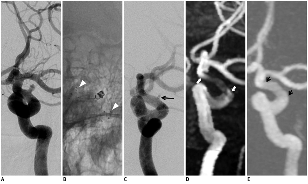
Fig. 2 53-year-old male with aneurysm at dorsal wall of distal internal carotid artery. A. Digital subtraction angiography shows saccular aneurysm at dorsal wall of C2 segment of left internal carotid artery. B. Neuroform stent (arrowheads) is inserted due to stretching of coil during embolization. C. Digital subtraction angiography immediately after stent-assisted coil embolization demonstrates residual neck (long arrow). D. 3D time of flight demonstrates moderate signal loss with focal artifactual luminal narrowing (open arrows) of stented segment and non-visualization of residual neck. E. 4D MR angiography with 1-day interval shows minimal signal loss of stented segment (short arrows) without luminal narrowing. Residual neck is not detected.
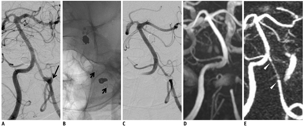
Fig. 3 59-year-old male with aneurysm at V4 segment of vertebral artery. A. Digital subtraction angiography shows broad-neck aneurysm (long arrow) at V4 segment of left vertebral artery involving posterior inferior cerebral artery origin. B. Stent-assisted coil embolization is successfully performed with Enterprise stent (short arrows). C. Digital subtraction angiography with 6-month interval shows patent stented segment and complete occlusion of treated aneurysm in left vertebral artery. D. 3D time of flight demonstrates complete signal loss causing non-visualization of stented segment of left vertebral artery. E. 4D MR angiography shows mild signal loss of stented segment and diffuse artifactual luminal narrowing (arrowheads). There is no evidence of recanalization of treated aneurysm.

Fig. 4 60-year-old male with large thrombosed aneurysm at vertebral artery. A. Digital subtraction angiography shows only non-thrombosed portion of large aneurysm. B. Stent-assisted coil embolization is performed using Neuroform stent (short arrows). C. Digital subtraction angiography after coil embolization shows minimal residual neck filling (long arrow). D. 3D time of flight after 1-day interval demonstrates diffuse narrowing of stented segment of left vertebral artery. However, it is difficult to evaluate completeness of coil embolization due to T1 shortening artifact of thrombosed aneurysm. E. 4D MR angiography clearly visualizes stented segment of left vertebral artery. Residual neck of aneurysm (arrowhead) is also suspected.
Cited by 2 articles
-
Time-of-Flight Magnetic Resonance Angiography for Follow-Up of Coil Embolization with Enterprise Stent for Intracranial Aneurysm: Usefulness of Source Images
Young Dae Cho, Kang Min Kim, Woong Jae Lee, Chul-Ho Sohn, Hyun-Seung Kang, Jeong Eun Kim, Moon Hee Han
Korean J Radiol. 2014;15(1):161-168. doi: 10.3348/kjr.2014.15.1.161.PulseRider Treated Aneurysm with Significant Artifact on Postoperative Magnetic Resonance Angiography: A Case Report and Literature Review
Anthony V. Nguyen, Laura K. Reed, Walter S. Lesley
Neurointervention. 2021;16(3):293-297. doi: 10.5469/neuroint.2021.00241.
Reference
-
1. Molyneux AJ, Kerr RS, Birks J, Ramzi N, Yarnold J, Sneade M, et al. Risk of recurrent subarachnoid haemorrhage, death, or dependence and standardised mortality ratios after clipping or coiling of an intracranial aneurysm in the International Subarachnoid Aneurysm Trial (ISAT): long-term follow-up. Lancet Neurol. 2009. 8:427–433.2. Wiebers DO, Whisnant JP, Huston J 3rd, Meissner I, Brown RD Jr, Piepgras DG, et al. Unruptured intracranial aneurysms: natural history, clinical outcome, and risks of surgical and endovascular treatment. Lancet. 2003. 362:103–110.3. Jabbour PM, Tjoumakaris SI, Rosenwasser RH. Endovascular management of intracranial aneurysms. Neurosurg Clin N Am. 2009. 20:383–398.4. Hwang JH, Roh HG, Chun YI, Kang HS, Choi JW, Moon WJ, et al. Endovascular coil embolization of very small intracranial aneurysms. Neuroradiology. 2011. 53:349–357.5. Chae KS, Jeon P, Kim KH, Kim ST, Kim HJ, Byun HS. Endovascular coil embolization of very small intracranial aneurysms. Korean J Radiol. 2010. 11:536–541.6. Tahtinen OI, Vanninen RL, Manninen HI, Rautio R, Haapanen A, Niskakangas T, et al. Wide-necked intracranial aneurysms: treatment with stent-assisted coil embolization during acute (<72 hours) subarachnoid hemorrhage--experience in 61 consecutive patients. Radiology. 2009. 253:199–208.7. Fiorella D, Albuquerque FC, Deshmukh VR, McDougall CG. Usefulness of the Neuroform stent for the treatment of cerebral aneurysms: results at initial (3-6-mo) follow-up. Neurosurgery. 2005. 56:1191–1201. discussion 1201-1192.8. Siddiqui MA, J JB, Lindsay KW, Jenkins S. Horizontal stent-assisted coil embolisation of wide-necked intracranial aneurysms with the Enterprise stent--a case series with early angiographic follow-up. Neuroradiology. 2009. 51:411–418.9. Kaufmann TJ, Huston J 3rd, Mandrekar JN, Schleck CD, Thielen KR, Kallmes DF. Complications of diagnostic cerebral angiography: evaluation of 19,826 consecutive patients. Radiology. 2007. 243:812–819.10. Schaafsma JD, Velthuis BK, Majoie CB, van den Berg R, Brouwer PA, Barkhof F, et al. Intracranial aneurysms treated with coil placement: test characteristics of follow-up MR angiography--multicenter study. Radiology. 2010. 256:209–218.11. Anzalone N, Scomazzoni F, Cirillo M, Righi C, Simionato F, Cadioli M, et al. Follow-up of coiled cerebral aneurysms at 3T: comparison of 3D time-of-flight MR angiography and contrast-enhanced MR angiography. AJNR Am J Neuroradiol. 2008. 29:1530–1536.12. Kwee TC, Kwee RM. MR angiography in the follow-up of intracranial aneurysms treated with Guglielmi detachable coils: systematic review and meta-analysis. Neuroradiology. 2007. 49:703–713.13. Majoie CB, Sprengers ME, van Rooij WJ, Lavini C, Sluzewski M, van Rijn JC, et al. MR angiography at 3T versus digital subtraction angiography in the follow-up of intracranial aneurysms treated with detachable coils. AJNR Am J Neuroradiol. 2005. 26:1349–1356.14. Pierot L, Delcourt C, Bouquigny F, Breidt D, Feuillet B, Lanoix O, et al. Follow-up of intracranial aneurysms selectively treated with coils: Prospective evaluation of contrast-enhanced MR angiography. AJNR Am J Neuroradiol. 2006. 27:744–749.15. Ramgren B, Siemund R, Cronqvist M, Undren P, Nilsson OG, Holtas S, et al. Follow-up of intracranial aneurysms treated with detachable coils: comparison of 3D inflow MRA at 3T and 1.5T and contrast-enhanced MRA at 3T with DSA. Neuroradiology. 2008. 50:947–995.16. Sprengers ME, Schaafsma JD, van Rooij WJ, van den Berg R, Rinkel GJ, Akkerman EM, et al. Evaluation of the occlusion status of coiled intracranial aneurysms with MR angiography at 3T: is contrast enhancement necessary? AJNR Am J Neuroradiol. 2009. 30:1665–1671.17. Yamada N, Hayashi K, Murao K, Higashi M, Iihara K. Time-of-flight MR angiography targeted to coiled intracranial aneurysms is more sensitive to residual flow than is digital subtraction angiography. AJNR Am J Neuroradiol. 2004. 25:1154–1157.18. Wall A, Kugel H, Bachman R, Matuszewski L, Kramer S, Heindel W, et al. 3.0 T vs. 1.5 T MR angiography: in vitro comparison of intravascular stent artifacts. J Magn Reson Imaging. 2005. 22:772–779.19. Wang Y, Truong TN, Yen C, Bilecen D, Watts R, Trost DW, et al. Quantitative evaluation of susceptibility and shielding effects of nitinol, platinum, cobalt-alloy, and stainless steel stents. Magn Reson Med. 2003. 49:972–976.20. Wallace RC, Karis JP, Partovi S, Fiorella D. Noninvasive imaging of treated cerebral aneurysms, part I: MR angiographic follow-up of coiled aneurysms. AJNR Am J Neuroradiol. 2007. 28:1001–1008.21. Cashen TA, Carr JC, Shin W, Walker MT, Futterer SF, Shaibani A, et al. Intracranial time-resolved contrast-enhanced MR angiography at 3T. AJNR Am J Neuroradiol. 2006. 27:822–829.22. Farb RI, Agid R, Willinsky RA, Johnstone DM, Terbrugge KG. Cranial dural arteriovenous fistula: diagnosis and classification with time-resolved MR angiography at 3T. AJNR Am J Neuroradiol. 2009. 30:1546–1551.23. Lim RP, Shapiro M, Wang EY, Law M, Babb JS, Rueff LE, et al. 3D time-resolved MR angiography (MRA) of the carotid arteries with time-resolved imaging with stochastic trajectories: comparison with 3D contrast-enhanced Bolus-Chase MRA and 3D time-of-flight MRA. AJNR Am J Neuroradiol. 2008. 29:1847–1854.24. Meckel S, Mekle R, Taschner C, Haller S, Scheffler K, Radue EW, et al. Time-resolved 3D contrast-enhanced MRA with GRAPPA on a 1.5-T system for imaging of craniocervical vascular disease: initial experience. Neuroradiology. 2006. 48:291–229.25. Tsuchiya K, Honya K, Fujikawa A, Tateishi H, Shiokawa Y. Postoperative assessment of extracranial-intracranial bypass by time-resolved 3D contrast-enhanced MR angiography using parallel imaging. AJNR Am J Neuroradiol. 2005. 26:2243–2247.26. Zou Z, Ma L, Cheng L, Cai Y, Meng X. Time-resolved contrast-enhanced MR angiography of intracranial lesions. J Magn Reson Imaging. 2008. 27:692–699.27. Roy D, Milot G, Raymond J. Endovascular treatment of unruptured aneurysms. Stroke. 2001. 32:1998–2004.28. Blum MB, Schmook M, Schernthaner R, Edelhauser G, Puchner S, Lammer J, et al. Quantification and detectability of instent stenosis with CT angiography and MR angiography in arterial stents in vitro. AJR Am J Roentgenol. 2007. 189:1238–1242.29. Lettau M, Sauer A, Heiland S, Rohde S, Bendszus M, Hahnel S. Carotid artery stents: in vitro comparison of different stent designs and sizes using CT angiography and contrast-enhanced MR angiography at 1.5T and 3T. AJNR Am J Neuroradiol. 2009. 30:1993–1997.30. Klisch J, Eger C, Sychra V, Strasilla C, Basche S, Weber J. Stent-assisted coil embolization of posterior circulation aneurysms using solitaire ab: preliminary experience. Neurosurgery. 2009. 65:258–266. discussion 266.31. Klisch J, Clajus C, Sychra V, Eger C, Strasilla C, Rosahl S, et al. Coil embolization of anterior circulation aneurysms supported by the Solitaire AB Neurovascular Remodeling Device. Neuroradiology. 2010. 52:349–359.
- Full Text Links
-
- Actions
-
Cited
- CITED
-
- Close
- Share
-
- Similar articles
-
- Selective Temporary Stent-Assisted Coil Embolization for Intracranial Wide-Necked Small Aneurysms Using Solitaire AB Retrievable Stent
- The Stent-Assisted Coil-Jailing Technique Facilitates Efficient Embolization of Tiny Cerebral Aneurysms
- Microcatheter-assisted Coil Embolization of Distal Vertebral Artery Wide-Necked Aneurysm: A Case Report
- A Complicated Case of Endovascular Stent Assisted Coil Embolization of an Aneurysm
- Stent-assisted Coil Embolization of Cerebral Aneurysms: Review Article
