Bone-added osteotome sinus floor elevation with simultaneous placement of non-submerged sand blasted with large grit and acid etched implants: a 5-year radiographic evaluation
- Affiliations
-
- 1Department of Periodontology, Yonsei University College of Dentistry, Seoul, Korea.
- 2Department of Periodontology, Research Institute for Periodontal Regeneration, Oral Science Research Center, Yonsei University College of Dentistry, Seoul, Korea. dentall@yuhs.ac
- KMID: 1783537
- DOI: http://doi.org/10.5051/jpis.2010.40.2.69
Abstract
- PURPOSE
Implant survival rates using a bone-added osteotome sinus floor elevation (BAOSFE) procedure with simultaneous placement of a non-submerged sand blasted with large grit and acid etched (SLA) implant are well documented at sites where native bone height is less than 5 mm. This study evaluated the clinical results of non-submerged SLA Straumann implants placed at the time of the BAOSFE procedure at sites where native bone height was less than 4 mm. Changes in graft height after the BAOSFE procedure were also assessed using radiographs for 5 years after the implant procedure.
METHODS
The BAOSFE procedure was performed on 4 patients with atrophic posterior maxillas with simultaneous placement of 7 non-submerged SLA implants. At least 7 standardized radiographs were obtained from each patient as follows: before surgery, immediately after implant placement, 6 months after surgery, every year for the next 3 years, and after more than 5 years had passed. Clinical and radiographic examinations were performed at every visit. Radiographic changes in graft height were calculated with respect to the implant's known length and the original sinus height.
RESULTS
All implants were stable functionally, as well as clinically and radiographically, during the follow-up. Most of the radiographic reduction in the grafted bone height occurred in the first 2 years; reduction after 2 years was slight.
CONCLUSIONS
The simultaneous placement of non-submerged SLA implants using the BAOSFE procedure is a feasible treatment option for patients with severe atrophic posterior maxillas. However, the grafted bone height is reduced during the healing period, and patients must be selected with care.
Keyword
MeSH Terms
Figure
-
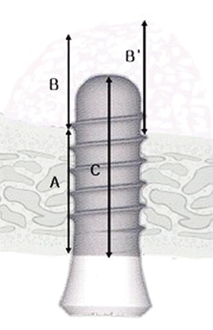
Figure 1 Schematic drawing of the measured parameters. (A) Native bone height: the distance from the alveolar crest to the floor of the maxillary sinus at the implant site, which was calculated as the mean of the mesial and distal native bone heights. Grafted bone height: the distance from the floor of the maxillary sinus to the border of the grafted bone at the implant site, which was calculated as the mean of the mesial (B) and distal (B') grafted bone heights. (C) The implant height: the distance from the apex to the head of the fixture.
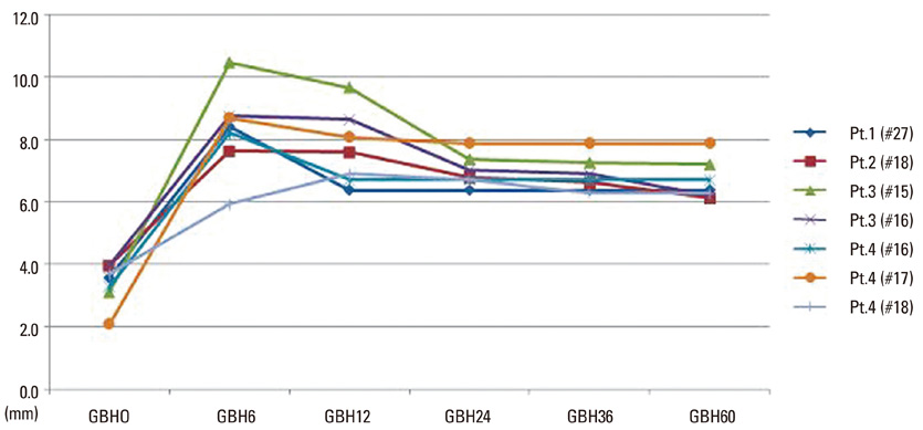
Figure 2 Periodical changes of grafted bone height based on the radiographic analysis. Results show that the notable radiographic changes occurred during the first 2 years after surgery. Numbers indicate the number of months elapsed since the surgery. Pt: patient, #: tooth number by F.D.I. numbering system, GBH: grafted bone height.
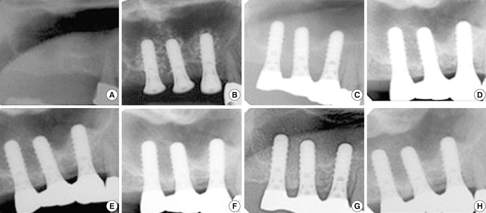
Figure 3 Radiographic evaluation of changes in grafted bone height using a series of radiographs from patient no. 4. The radiographs show remodeling of the grafted bone during the first post-surgery examination. (A) Prior to surgery, (B) immediately after surgery, (C) six months after surgery, (D) one year after surgery, (E) two years after surgery, (F) three years after surgery, (G) five years after surgery, (H) six years after surgery.
Reference
-
1. Jensen J, Simonsen EK, Sindet-Pedersen S. Reconstruction of the severely resorbed maxilla with bone grafting and osseointegrated implants: a preliminary report. J Oral Maxillofac Surg. 1990. 48:27–32.
Article2. Adell R, Lekholm U, Grondahl K, Branemark PI, Lindstrom J, Jacobsson M. Reconstruction of severely resorbed edentulous maxillae using osseointegrated fixtures in immediate autogenous bone grafts. Int J Oral Maxillofac Implants. 1990. 5:233–246.3. Isaksson S. Evaluation of three bone grafting techniques for severely resorbed maxillae in conjunction with immediate endosseous implants. Int J Oral Maxillofac Implants. 1994. 9:679–688.4. Kahnberg KE, Nystrom E, Bartholdsson L. Combined use of bone grafts and Branemark fixtures in the treatment of severely resorbed maxillae. Int J Oral Maxillofac Implants. 1989. 4:297–304.5. Fugazzotto PA. Maxillary sinus grafting with and without simultaneous implant placement: technical considerations and case reports. Int J Periodontics Restorative Dent. 1994. 14:544–551.6. Fugazzotto PA, Vlassis J. Long-term success of sinus augmentation using various surgical approaches and grafting materials. Int J Oral Maxillofac Implants. 1998. 13:52–58.7. Blomqvist JE, Alberius P, Isaksson S. Two-stage maxillary sinus reconstruction with endosseous implants: a prospectivestudy. Int J Oral Maxillofac Implants. 1998. 13:758–766.8. Summers RB. A new concept in maxillary implant surgery: the osteotome technique. Compendium. 1994. 15:152–158.9. Summers RB. The osteotome technique: Part 3--Less invasive methods of elevating the sinus floor. Compendium. 1994. 15:698–704.10. Zitzmann NU, Scharer P. Sinus elevation procedures in the resorbed posterior maxilla: comparison of the crestal and lateral approaches. Oral Surg Oral Med Oral Pathol Oral Radiol Endod. 1998. 85:8–17.11. Rosen PS, Summers R, Mellado JR, Salkin LM, Shanaman RH, Marks MH, et al. The bone-added osteotome sinus floor elevation technique: multicenter retrospective report of consecutively treated patients. Int J Oral Maxillofac Implants. 1999. 14:853–858.12. Bruschi GB, Scipioni A, Calesini G, Bruschi E. Localized management of sinus floor with simultaneous implant placement: a clinical report. Int J Oral Maxillofac Implants. 1998. 13:219–226.13. Winter AA, Pollack AS, Odrich RB. Placement of implants in the severely atrophic posterior maxilla using localized management of the sinus floor: a preliminary study. Int J Oral Maxillofac Implants. 2002. 17:687–695.14. Horowitz RA. The use of osteotomes for sinus augmentation at the time of implant placement. Compend Contin Educ Dent. 1997. 18:441–452.15. Komarnyckyj OG, London RM. Osteotome single-stage dental implant placement with and without sinus elevation: a clinical report. Int J Oral Maxillofac Implants. 1998. 13:799–804.16. Tong DC, Rioux K, Drangsholt M, Beirne OR. A review of survival rates for implants placed in grafted maxillary sinuses using meta-analysis. Int J Oral Maxillofac Implants. 1998. 13:175–182.17. Ferrigno N, Laureti M, Fanali S. Dental implants placement in conjunction with osteotome sinus floor elevation: a 12-year life-table analysis from a prospective studyon 588 ITI implants. Clin Oral Implants Res. 2006. 17:194–205.
Article18. Diserens V, Mericske E, Schappi P, Mericske-Stern R. Transcrestal sinus floor elevation: report of a case series. Int J Periodontics Restorative Dent. 2006. 26:151–159.19. Wennerberg A, Albrektsson T, Johansson C, Andersson B. Experimental study of turned and grit-blasted screw-shaped implants with special emphasis on effects of blasting material and surface topography. Biomaterials. 1996. 17:15–22.
Article20. Ogawa T, Ozawa S, Shih JH, Ryu KH, Sukotjo C, Yang JM, et al. Biomechanical evaluation of osseous implants having different surface topographies in rats. J Dent Res. 2000. 79:1857–1863.
Article21. Pinholt EM. Branemark and ITI dental implants in the human bone-grafted maxilla: a comparative evaluation. Clin Oral Implants Res. 2003. 14:584–592.
Article22. Cavicchia F, Bravi F, Petrelli G. Localized augmentation of the maxillary sinus floor through a coronal approach for the placement of implants. Int J Periodontics Restorative Dent. 2001. 21:475–485.23. Fugazzotto PA. Augmentation of the posterior maxilla: a proposed hierarchy of treatment selection. J Periodontol. 2003. 74:1682–1691.
Article24. Peleg M, Mazor Z, Garg AK. Augmentation grafting of the maxillary sinus and simultaneous implant placement in patients with 3 to 5 mm of residual alveolar bone height. Int J Oral Maxillofac Implants. 1999. 14:549–556.25. Buchmann R, Khoury F, Faust C, Lange DE. Peri-implant conditions in periodontally compromised patients following maxillary sinus augmentation: a long-term posttherapy trial. Clin Oral Implants Res. 1999. 10:103–110.
Article26. Raghoebar GM, Timmenga NM, Reintsema H, Stegenga B, Vissink A. Maxillary bone grafting for insertion of endosseous implants: results after 12-124 months. Clin Oral Implants Res. 2001. 12:279–286.
Article27. Hallman M, Hedin M, Sennerby L, Lundgren S. A prospective 1-year clinical and radiographic study of implants placed after maxillary sinus floor augmentation with bovine hydroxyapatite and autogenous bone. J Oral Maxillofac Surg. 2002. 60:277–284.
Article28. Geurs NC, Wang IC, Shulman LB, Jeffcoat MK. Retrospective radiographic analysis of sinus graft and implant placement procedures from the Academy of Osseointegration Consensus Conference on Sinus Grafts. Int J Periodontics Restorative Dent. 2001. 21:517–523.29. Bragger U, Gerber C, Joss A, Haenni S, Meier A, Hashorva E, et al. Patterns of tissue remodeling after placement of ITI dental implants using an osteotome technique: a longitudinal radiographic case cohort study. Clin Oral Implants Res. 2004. 15:158–166.
Article30. Diserens V, Mericske E, Mericske-Stern R. Radiographic analysis of the transcrestal sinus floor elevation: short-term observations. Clin Implant Dent Relat Res. 2005. 7:70–78.
Article31. Hatano N, Shimizu Y, Ooya K. A clinical long-term radiographic evaluation of graft height changes after maxillary sinus floor augmentation with a 2:1 autogenous bone/xenograft mixture and simultaneous placement of dental implants. Clin Oral Implants Res. 2004. 15:339–345.
Article
- Full Text Links
-
- Actions
-
Cited
- CITED
-
- Close
- Share
-
- Similar articles
-
- Scanning Electron Microscopic Study of the Effects of Citric Acid on the Change of Implant Surface According to Application Time
- Endosinus Bone Gain after Osteotome Sinus Floor Elevation Without Bone Grafting: A Retrospective Study
- Implantation Using Osteotome Sinus Floor Elevation Procedure
- Resorption of bone graft after maxillary sinus grafting and simultaneous implant placement
- Clinical evaluation of Branemark Ti-Unite implant and ITI SLA implant in the post maxillary area with sinus elevation technique
