J Korean Assoc Oral Maxillofac Surg.
2014 Jun;40(3):117-122.
Resorption of bone graft after maxillary sinus grafting and simultaneous implant placement
- Affiliations
-
- 1Department of Oral and Maxillofacial Surgery, Section of Dentistry, Seoul National University Bundang Hospital, Seongnam, Korea.
- 2Department of Oral and Maxillofacial Surgery, School of Dentistry, Chosun University, Gwangju, Korea. sgckim@chosun.ac.kr
Abstract
OBJECTIVES
The purpose of this study was to evaluate the sinus bone graft resorption over 3 years after two-stage implant placement.
MATERIALS AND METHODS
The subjects for this study included 30 patients whose maxillary posterior ridges were too atrophic for implants. Bone-added osteotome sinus floor elevation was used in 15 maxillary sinuses, while the bone graft by lateral approach technique was used in 25 maxillary sinuses. The height from the top of the fixture to the sinus floor was estimated immediately after implant placement and the follow-up period was over 3 years. The surgery was classified with two groups: sinus bone grafting with and without autogenous bone. All implants were placed simultaneously.
RESULTS
The mean vertical bone loss was 3.15+/-2.95 mm. The survival rate of implants was 94.7%.
CONCLUSION
The amount of bone resorption was not significantly associated with the surgical methods, the type of bone graft materials used, or sinus perforation during surgery.
MeSH Terms
Figure
-
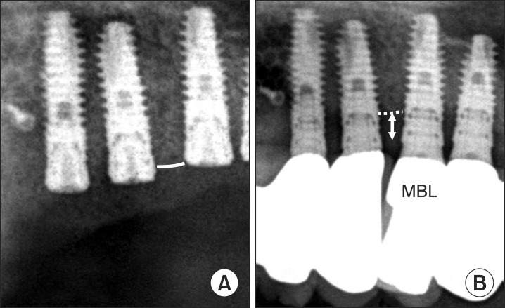
Fig. 1 Sinus bone resorption at long-term follow-up. The amount of resorption is defined as subraction of the final graft height (FGH) from the initial graft height (IGH). A. Right after sinus bone graft. B. At final follow-up.

Fig. 2 Marginal bone loss (MBL) measurement. A. Right after sinus bone graft. B. At final follow-up.
Reference
-
1. Hatano N, Shimizu Y, Ooya K. A clinical long-term radiographic evaluation of graft height changes after maxillary sinus floor augmentation with a 2:1 autogenous bone/xenograft mixture and simultaneous placement of dental implants. Clin Oral Implants Res. 2004; 15:339–345. PMID: 15142097.2. Zijderveld SA, Schulten EA, Aartman IH, ten Bruggenkate CM. Long-term changes in graft height after maxillary sinus floor elevation with different grafting materials: radiographic evaluation with a minimum follow-up of 4.5 years. Clin Oral Implants Res. 2009; 20:691–700. PMID: 19453567.
Article3. Rosen PS, Summers R, Mellado JR, Salkin LM, Shanaman RH, Marks MH, et al. The bone-added osteotome sinus floor elevation technique: multicenter retrospective report of consecutively treated patients. Int J Oral Maxillofac Implants. 1999; 14:853–858. PMID: 10612923.4. Summers RB. Sinus floor elevation with osteotomes. J Esthet Dent. 1998; 10:164–171. PMID: 9759033.
Article5. Kim YK, Yun PY, Kim SG, Kim BS, Ong JL. Evaluation of sinus bone resorption and marginal bone loss after sinus bone grafting and implant placement. Oral Surg Oral Med Oral Pathol Oral Radiol Endod. 2009; 107:e21–e28. PMID: 19138634.
Article6. Kim YK, Yun PY, Kim SG, Lim SC. Analysis of the healing process in sinus bone grafting using various grafting materials. Oral Surg Oral Med Oral Pathol Oral Radiol Endod. 2009; 107:204–211. PMID: 18801669.
Article7. Block MS, Kent JN, Kallukaran FU, Thunthy K, Weinberg R. Bone maintenance 5 to 10 years after sinus grafting. J Oral Maxillofac Surg. 1998; 56:706–714. PMID: 9632328.
Article8. Yildirim M, Spiekermann H, Biesterfeld S, Edelhoff D. Maxillary sinus augmentation using xenogenic bone substitute material Bio-Oss in combination with venous blood. A histologic and histomorphometric study in humans. Clin Oral Implants Res. 2000; 11:217–229. PMID: 11168213.9. Mayfield LJ, Skoglund A, Hising P, Lang NP, Attström R. Evaluation following functional loading of titanium fixtures placed in ridges augmented by deproteinized bone mineral. A human case study. Clin Oral Implants Res. 2001; 12:508–514. PMID: 11564112.10. Piattelli M, Favero GA, Scarano A, Orsini G, Piattelli A. Bone reactions to anorganic bovine bone (Bio-Oss) used in sinus augmentation procedures: a histologic long-term report of 20 cases in humans. Int J Oral Maxillofac Implants. 1999; 14:835–840. PMID: 10612920.11. Hallman M, Cederlund A, Lindskog S, Lundgren S, Sennerby L. A clinical histologic study of bovine hydroxyapatite in combination with autogenous bone and fibrin glue for maxillary sinus floor augmentation. Results after 6 to 8 months of healing. Clin Oral Implants Res. 2001; 12:135–143. PMID: 11251663.12. Gosain AK. Hydroxyapatite cement paste cranioplasty for the treatment of temporal hollowing after cranial vault remodeling in a growing child. J Craniofac Surg. 1997; 8:506–511. PMID: 9477838.
Article13. Asai S, Shimizu Y, Ooya K. Maxillary sinus augmentation model in rabbits: effect of occluded nasal ostium on new bone formation. Clin Oral Implants Res. 2002; 13:405–409. PMID: 12175378.
Article14. Artzi Z, Weinreb M, Givol N, Rohrer MD, Nemcovsky CE, Prasad HS, et al. Biomaterial resorption rate and healing site morphology of inorganic bovine bone and beta-tricalcium phosphate in the canine: a 24-month longitudinal histologic study and morphometric analysis. Int J Oral Maxillofac Implants. 2004; 19:357–368. PMID: 15214219.15. Aaboe M, Pinholt EM, Hjørting-Hansen E. Healing of experimentally created defects: a review. Br J Oral Maxillofac Surg. 1995; 33:312–318. PMID: 8555150.
Article16. Chanavaz M. Maxillary sinus: anatomy, physiology, surgery, and bone grafting related to implantology--eleven years of surgical experience (1979-1990). J Oral Implantol. 1990; 16:199–209. PMID: 2098563.17. Hürzeler MB, Kirsch A, Ackermann KL, Quiñones CR. Reconstruction of the severely resorbed maxilla with dental implants in the augmented maxillary sinus: a 5-year clinical investigation. Int J Oral Maxillofac Implants. 1996; 11:466–475. PMID: 8803342.18. Nyström E, Kahnberg KE, Albrektsson T. Treatment of the severely resorbed maxillae with bone graft and titanium implants: histologic review of autopsy specimens. Int J Oral Maxillofac Implants. 1993; 8:167–172. PMID: 8359872.19. Keller EE, Eckert SE, Tolman DE. Maxillary antral and nasal one-stage inlay composite bone graft: preliminary report on 30 recipient sites. J Oral Maxillofac Surg. 1994; 52:438–447. PMID: 8169704.
Article20. Listrom RD, Symington JM. Osseointegrated dental implants in conjunction with bone grafts. Int J Oral Maxillofac Surg. 1988; 17:116–118. PMID: 3133419.
Article21. Bravetti P, Membre H, Marchal L, Jankowski R. Histologic changes in the sinus membrane after maxillary sinus augmentation in goats. J Oral Maxillofac Surg. 1998; 56:1170–1176. PMID: 9766543.
Article22. Mills MP. Spontaneous early exposure of submerged endosseous implants resulting in crestal bone loss: a clinical evaluation between stage I and stage II surgery. Implant Dent. 2003; 12:9–10.23. Wang CY, Stashenko P. The role of interleukin-1 alpha in the pathogenesis of periapical bone destruction in a rat model system. Oral Microbiol Immunol. 1993; 8:50–56. PMID: 8510985.
- Full Text Links
-
- Actions
-
Cited
- CITED
-
- Close
- Share
-
- Similar articles
-
- Clinical Study on Implant Survival and Graft Resorption Rate After Maxillary Sinus Bone Grafting
- One-staged sinus bone graft using tapered porous surfaced implant in lower residual bone height of maxillary sinus
- Assessment of the autogenous bone graft for sinus elevation
- Maxillary Sinus Augmentation Using Autogenous Teeth: Preliminary Report
- Implant Surgery with Both Sinus Bone Graft in the Maxillary edentulous patient: A Case Report
