Analysis of location and prevalence of maxillary sinus septa
- Affiliations
-
- 1Department of Periodontology, Chonbuk National University School of Dentistry, Jeonju, Korea. cbuperio@chonbuk.ac.kr
- 2Research Institute of Oral Bio-Science, Chonbuk National University, Jeonju, Korea.
- KMID: 1783535
- DOI: http://doi.org/10.5051/jpis.2010.40.2.56
Abstract
- PURPOSE
The sinus lift procedure requires detailed knowledge of maxillary sinus anatomy and the possible anatomical variations. This study evaluated the location and prevalence of maxillary sinus septa using computed tomography (CT).
METHODS
This study was based on the analysis of CT images for posterior maxilla which were obtained from patients who visited Chonbuk National University Dental Hospital during the period of June 2007 to December 2008. With the exclusion of cases presenting any pathological changes, 236 maxillary sinuses in 204 patients were retrospectively analyzed. The average age of the patients was 50.9. The cases were divided into two groups, an atrophy/edentulous segment and a non-atrophy/dentate segment, and maxillary sinus septa of less than 2.5 mm were not taken in-to consideration. The location of septa was also divided for analysis into 3 regions: the anterior (1st and 2nd premolar), middle (1st and 2nd molar) and posterior (behind 2nd molar) regions.
RESULTS
In 54 (20.9%) of the 204 patients there were pathologic findings, and those patients were excluded from the analysis. Sinus septa were present in 58 (24.6%) of the 236 maxillary sinuses and in 55 (27%) of the 204 total patients. In the atrophy/edentulous ridge group (148 maxillary sinuses), 41 cases (27.7%) were found, and 17 cases (19.3%) were found in the non-atrophy/dentulous ridge group (88 maxillary sinuses). In terms of location, septa were found in 18 cases (27.3%) in the anterior, in 33 cases (50%) in the middle and in 15 cases (22.7%) in the posterior regions.
CONCLUSIONS
In the posterior maxilla, regardless of type of ridge (atrophy/edentulous or non-atrophy/dentate), the anatomical variation of sinus septa is diverse in its prevalence and location. Thus, accurate information on the maxillary sinus of the patient is essential and should be clearly understood by the surgeon to prevent possible complications during sinus lifting.
MeSH Terms
Figure
-
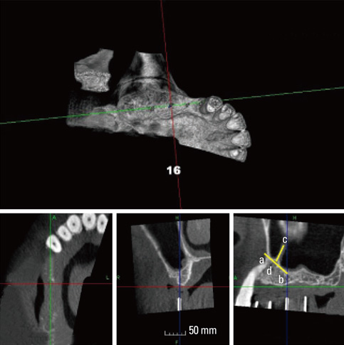
Figure 1 Evaluation of septa at mid-crestal aspect (lower-right). A line drawn at the approximate base of the septa was established (A, B), and its height was measured using a line extending from this base to the most coronal portion of the septa (C, D). If the vertical dimension of the septa was over 2.5 mm, the septa was included in our study.
Cited by 2 articles
-
Frequency of different maxillary sinus septal patterns found on cone-beam computed tomography and predicting the associated risk of sinus membrane perforation during sinus lifting
Ali Khalighi Sigaroudi, Zahra Dalili Kajan, Shabnam Rastgar, Hamid Neshandar Asli
Imaging Sci Dent. 2017;47(4):261-267. doi: 10.5624/isd.2017.47.4.261.The incidence and morphology of maxillary sinus septa in dentate and edentulous maxillae: a cadaveric study with a brief review of the literature
Kusum Rajendra Gandhi, Rajendra Namdeo Wabale, Abu Ubaida Siddiqui, Mujjebuddeen Samsudeen Farooqui
J Korean Assoc Oral Maxillofac Surg. 2015;41(1):30-36. doi: 10.5125/jkaoms.2015.41.1.30.
Reference
-
1. Chanavaz M. Maxillary sinus: anatomy, physiology, surgery, and bone grafting related to implantology--eleven years of surgical experience (1979-1990). J Oral Implantol. 1990. 16:199–209.2. Cawood JI, Howell RA. A classification of the edentulous jaws. Int J Oral Maxillofac Surg. 1988. 17:232–236.
Article3. Vinter I, Krmpotic-Nemanic J, Hat J, Jalsovec D. Does the alveolar process of the maxilla always disappear after tooth loss? Laryngorhinootologie. 1993. 72:605–607.4. Underwood AS. An inquiry into the anatomy and pathology of the maxillary sinus. J Anat Physiol. 1910. 44:354–369.5. Boyne PJ, James RA. Grafting of the maxillary sinus floor with autogenous marrow and bone. J Oral Surg. 1980. 38:613–616.6. Farmand M. Horse-shoe sandwich osteotomy of the edentulous maxilla as a preprosthetic procedure. J Maxillofac Surg. 1986. 14:238–244.
Article7. Sailer HF. A new method of inserting endosseous implants in totally atrophic maxillae. J Craniomaxillofac Surg. 1989. 17:299–305.
Article8. Beaumont C, Zafiropoulos GG, Rohmann K, Tatakis DN. Prevalence of maxillary sinus disease and abnormalities in patients scheduled for sinus lift procedures. J Periodontol. 2005. 76:461–467.
Article9. Kasabah S, Slezak R, Simunek A, Krug J, Lecaro MC. Evaluation of the accuracy of panoramic radiograph in the definition of maxillary sinus septa. Acta Medica (Hradec Kralove). 2002. 45:173–175.
Article10. Maksoud MA. Complications after maxillary sinus augmentation: a case report. Implant Dent. 2001. 10:168–171.
Article11. Ueda M, Kaneda T. Maxillary sinusitis caused by dental implants: report of two cases. J Oral Maxillofac Surg. 1992. 50:285–287.
Article12. Ulm CW, Solar P, Krennmair G, Matejka M, Watzek G. Incidence and suggested surgical management of septa in sinus-lift procedures. Int J Oral Maxillofac Implants. 1995. 10:462–465.13. Tatum H Jr. Maxillary and sinus implant reconstructions. Dent Clin North Am. 1986. 30:207–229.14. Betts NJ, Miloro M. Modification of the sinus lift procedure for septa in the maxillary antrum. J Oral Maxillofac Surg. 1994. 52:332–333.
Article15. van den Bergh JP, ten Bruggenkate CM, Disch FJ, Tuinzing DB. Anatomical aspects of sinus floor elevations. Clin Oral Implants Res. 2000. 11:256–265.
Article16. Krennmair G, Ulm CW, Lugmayr H, Solar P. The incidence, location, and height of maxillary sinus septa in the edentulous and dentate maxilla. J Oral Maxillofac Surg. 1999. 57:667–671.
Article17. Neivert H. Symposium on maxillary sinus: surgical anatomy of the maxillary sinus. Laryngoscope. 1930. 40:1–4.18. Tidwell JK, Blijdorp PA, Stoelinga PJ, Brouns JB, Hinderks F. Composite grafting of the maxillary sinus for placement of endosteal implants: a preliminary report of 48 patients. Int J Oral Maxillofac Surg. 1992. 21:204–209.
Article19. Kim MJ, Jung UW, Kim CS, Kim KD, Choi SH, Kim CK, et al. Maxillary sinus septa: prevalence, height, location, and morphology. A reformatted computed tomography scan analysis. J Periodontol. 2006. 77:903–908.
Article20. Velasquez-Plata D, Hovey LR, Peach CC, Alder ME. Maxillary sinus septa: a 3-dimensional computerized tomographic scan analysis. Int J Oral Maxillofac Implants. 2002. 17:854–860.21. Krennmair G, Ulm C, Lugmayr H. Maxillary sinus septa: incidence, morphology and clinical implications. J Craniomaxillofac Surg. 1997. 25:261–265.
Article
- Full Text Links
-
- Actions
-
Cited
- CITED
-
- Close
- Share
-
- Similar articles
-
- Maxillary sinus septa: comparison between panoramic radiography and CBCT
- CLINICO-ANATOMICAL STUDY OF SEPTUM IN THE MAXILLARY SINUS
- The clinical study of maxillary sinus septa used in panorama, CT
- The incidence and morphology of maxillary sinus septa in dentate and edentulous maxillae: a cadaveric study with a brief review of the literature
- Morphological analysis of maxillary sinus septum using computed tomography
