The incidence and morphology of maxillary sinus septa in dentate and edentulous maxillae: a cadaveric study with a brief review of the literature
- Affiliations
-
- 1Department of Anatomy, Rural Medical College, Pravara Institute of Medical Sciences, Loni, Maharashtra, India. gandhikusum.r@gmail.com
- 2All India Institute of Medical Sciences, Tatibandh, Raipur, Chattisgarh, India.
- KMID: 2328651
- DOI: http://doi.org/10.5125/jkaoms.2015.41.1.30
Abstract
OBJECTIVES
The aim of this study is to determine the incidence, location, and orientation of maxillary sinus septa in formalin embalmed cadavers.
MATERIALS AND METHODS
The study was conducted on 210 cadaveric heads available in our department. After taking the mid-sagittal section the specimens were opened from the medial aspect and the sinus cavity was explored for the presence of maxillary sinus septa, their anatomical plane, location and dimensions.
RESULTS
The mean linear distance between maxillary sinus floor and its anatomical ostium was 26.76+/-5.21 mm and 26.91+/-4.96 mm on right and left side, respectively. A total of 59 maxillary sinus septa (28.1%) were observed in 210 maxillary specimens. Septae were most common, 33 septa (55.9%), in the middle region (between first and second molar tooth) of the sinus cavity. The maxillary sinus membrane (Schneiderian membrane) adhered tightly to the maxillary sinus and over the septae. Significantly more maxillary sinus septa were observed in edentulous maxillae in comparison to the dentate upper jaw.
CONCLUSION
Knowledge of location of maxillary sinus ostium is mandatory for the rhinologist for drainage of secretions in maxillary sinusitis. The morphological details of maxillary sinus septa, particularly their location and anatomical planes, will guide dentists in performance of safe implant surgeries. The maxillary antrum septa of category I and II may complicate the procedure of inversion of bone plate and elevation of sinus membrane during maxillary augmentation surgeries. The category III septa observed in the sagittal plane were embedded by one of the branches of the infraorbital nerve in it, and if accidentally cut will lead to infraorbital nerve palsy in maxillary sinus surgeries.
MeSH Terms
Figure
-
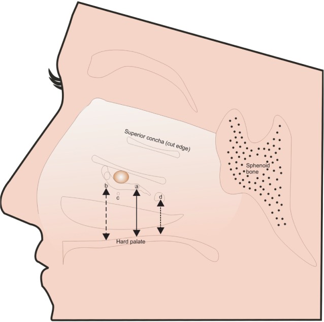
Fig. 1 The schematic diagram of the lateral nasal wall where the three conchae has been removed to show the distance between nasal floor and maxillary sinus ostium. 'a' denote the normal anatomical location of maxillary ostium in posterior part of semilunar hiatus. 'b, c, and d' shows the location of maxillary ostium inferior and posterior to semilunar hiatus.
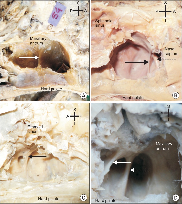
Fig. 2 The location and orientation of maxillary sinus septa (MSS) in various anatomical planes. A. The MSS arising from floor of right maxillary sinus and located between the first and second molar tooth in the middle region (arrow). Septa is dividing the maxillary antrum into two incomplete (antrium and posterior) compartments. B. The MSS arising from lateral wall of left maxillary autrum shown with solid arrow. Note the ransverse orientation of the septa. This septa has largest dimension. The infraorbital nerve is piercing the MSS shown with dotted arrow. C. Single incomplete septa located in antero-superior quadrant of right maxillary autrum (arrow). The anterior superior alveolar nerve is embedded within the septa. D. Double septa is observed in the antero-superior portion of antrum on left side and both the septa are occupied by branches of infra orbital nerve (solid and dotted arrows). (S: superior, I: inferior, A: anterior, P: posterior)
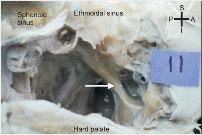
Fig. 3 Infraorbital nerve is traversing in a thin bony trabeculli in the superior part of left maxillary antrum (arrow). (S: superior, I: inferior, A: anterior, P: posterior)
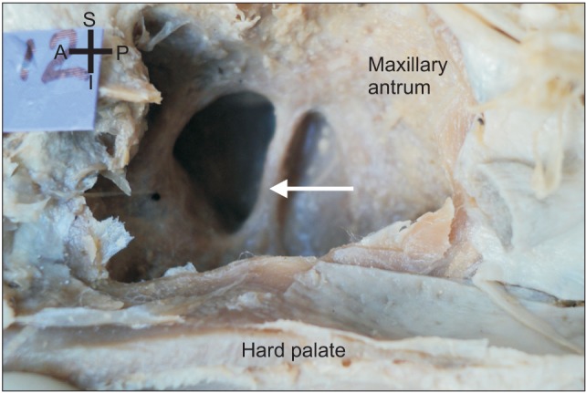
Fig. 4 Middle superior alveolar nerve is crossing in the center of the maxillary antrum in a separate bony canal (arrow). (S: superior, I: inferior, A: anterior, P: posterior)
Reference
-
1. Woo I, Le BT. Maxillary sinus floor elevation: review of anatomy and two techniques. Implant Dent. 2004; 13:28–32. PMID: 15017301.
Article2. Rosano G, Taschieri S, Gaudy JF, Lesmes D, Del Fabbro M. Maxillary sinus septa: a cadaveric study. J Oral Maxillofac Surg. 2010; 68:1360–1364. PMID: 20231050.
Article3. Pikos MA. Maxillary sinus membrane repair: update on technique for large and complete perforations. Implant Dent. 2008; 17:24–31. PMID: 18332755.
Article4. Underwood AS. An inquiry into the anatomy and pathology of the maxillary sinus. J Anat Physiol. 1910; 44:354–369. PMID: 17232856.5. Kim MJ, Jung UW, Kim CS, Kim KD, Choi SH, Kim CK, et al. Maxillary sinus septa: prevalence, height, location, and morphology. A reformatted computed tomography scan analysis. J Periodontol. 2006; 77:903–908. PMID: 16671885.
Article6. Maestre-Ferrín L, Galán-Gil S, Rubio-Serrano M, Peñarrocha-Diago M, Peñarrocha-Oltra D. Maxillary sinus septa: a systematic review. Med Oral Patol Oral Cir Bucal. 2010; 15:e383–e386. PMID: 19767706.7. Uchida Y, Goto M, Katsuki T, Akiyoshi T. A cadaveric study of maxillary sinus size as an aid in bone grafting of the maxillary sinus floor. J Oral Maxillofac Surg. 1998; 56:1158–1163. PMID: 9766541.
Article8. Gosau M, Rink D, Driemel O, Draenert FG. Maxillary sinus anatomy: a cadaveric study with clinical implications. Anat Rec (Hoboken). 2009; 292:352–354. PMID: 19248167.
Article9. May M, Sobol SM, Korzec K. The location of the maxillary os and its importance to the endoscopic sinus surgeon. Laryngoscope. 1990; 100:1037–1042. PMID: 2215032.
Article10. Shibli JA, Faveri M, Ferrari DS, Melo L, Garcia RV, d'Avila S, et al. Prevalence of maxillary sinus septa in 1024 subjects with edentulous upper jaws: a retrospective study. J Oral Implantol. 2007; 33:293–296. PMID: 17987862.
Article11. Lee WJ, Lee SJ, Kim HS. Analysis of location and prevalence of maxillary sinus septa. J Periodontal Implant Sci. 2010; 40:56–60. PMID: 20498761.
Article12. Jung JW, Song KH, Lee SK, Kim JY, Yang BE, Kim SG, et al. The clinical study of maxillary sinus septa used in panorama, CT. J Korean Assoc Oral Maxillofac Surg. 2008; 34:319–324.13. Mailleux P, Desgain O, Ingabire MI. Ectopic infraorbital nerve in a maxillary sinus septum: another potentially dangerous variant for sinus surgery. JBR-BTR. 2010; 93:308–309. PMID: 21381529.
Article14. Oh E, Kraut RA. Effect of sinus membrane perforation on dental implant integration: a retrospective study on 128 patients. Implant Dent. 2011; 20:13–19. PMID: 21278522.
Article15. Chanavaz M. Maxillary sinus: anatomy, physiology, surgery, and bone grafting related to implantology--eleven years of surgical experience (1979-1990). J Oral Implantol. 1990; 16:199–209. PMID: 2098563.16. Neivert H. Symposium on maxillary sinus: surgical anatomy of the maxillary sinus. Laryngoscope. 1930; 40:1–4.
- Full Text Links
-
- Actions
-
Cited
- CITED
-
- Close
- Share
-
- Similar articles
-
- The clinical study of maxillary sinus septa used in panorama, CT
- Analysis of location and prevalence of maxillary sinus septa
- CLINICO-ANATOMICAL STUDY OF SEPTUM IN THE MAXILLARY SINUS
- Maxillary sinus septa: comparison between panoramic radiography and CBCT
- A Retrospective study on the survival rate of the sinus perforated implants
