J Korean Soc Magn Reson Med.
2010 Jun;14(1):56-63. 10.13104/jksmrm.2010.14.1.56.
Cortical Thickness Estimation Using DIR Imaging with GRAPPA Factor 2
- Affiliations
-
- 1School of Electrical and Electronic Engineering, Yonsei University, Korea. donghyunkim@yonsei.ac.kr
- 2Department of Radiology, Yonsei University, Korea.
- KMID: 1782994
- DOI: http://doi.org/10.13104/jksmrm.2010.14.1.56
Abstract
- PURPOSE
DIR image is relatively free from susceptibility artifacts therefore, DIR image can make it possible to reliably measure cortical thickness/volume. One drawback of the DIR acquisition is the long scan time to acquire the fully sampled 3D data set. To solve this problem, we applied a parallel imaging method (GRAPPA) and verify the reliability of using the volumetric study.
MATERIALS AND METHODS
Six healthy volunteers (3 males and 3 females; age 25.33+/-2.25 years) underwent MRI using the 3D DIR sequence at a 3.0T Siemens Tim Trio MRI scanner. GRAPPA simulation was performed from the fully sampled data set for reduction factor 2. Data reconstruction was performed using MATLAB R2009b. Freesurfer v.4.3.0 was used to evaluate the cortical thickness of the entire brain, and to extract white matter information from the DIR image, Analyze 9.0 was used. The global cortical thickness estimated from the reconstructed image was compared with reference image by using a T-test in SPSS.
RESULTS
Although reduced SNR and blurring are observed from the reconstructed image, in terms of segmentation the effect was not so significant. The volumetric result was validated that there were no significant differences in many cortical regions.
CONCLUSION
This study was performed with DIR image for a volumetric MRI study. To solve the long scan time of 3D DIR imaging, we applied GRAPPA algorithm. According to the results, fast imaging can be done with reduction factor 2 with little loss of image quality at 3.0T.
Keyword
Figure
-

Fig. 1

Fig. 2 simple pulse diagram.

Fig. 3

Fig. 4

Fig. 5

Fig. 6
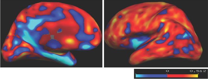
Fig. 7

Fig. 8
Reference
-
1. Mugler JP 3rd, Brookeman JR. Rapid three-dimensional T1-weighted MR imaging with the MP-RAGE sequence. J Magn Reson Imaging. 1991. 1(5):561–567.2. Pouwels PJ, Kuijer JP, Mugler JP, 3rd , Guttmann CR, Barkhof F. Human gray matter: feasibility of single-slab 3D double inversion-recovery high-spatial-resolution MR imaging. Radiology. 2006. 241(3):873–879.3. Yoo E, Kim DH, Park HJ, Kim EY. Unreliability of Cortical Volumetry in Regions near The Skull Base on 3D T1-weighted Imaging: Comparision study with 3D Double Inversion-recovery Imaging. 2008. In : 16th Scientific Meeting of the International Society of Magnetic Resonance in Medicine; Toronto, Canada. 3502.4. Edelman RR, Chien D, Kim D. Fast selective black blood MR imaging. Radiology. 1991. 181(3):655–660.5. Griswold MA, Jakob PM, Heidemann RM, Nittka M, Jellus V, Wang J, Kiefer B, Haase A. Generalized autocalibrating partially parallel acquisitions (GRAPPA). Magn Reson Med. 2002. 47(6):1202–1210.6. Pruessmann KP, Weiger M, Scheidegger MB, Boesiger P. SENSE: sensitivity encoding for fast MRI. Magn Reson Med. 1999. 42(5):952–962.7. Sodickson DK, Manning WJ. Simultaneous acquisition of spatial harmonics (SMASH): fast imaging with radiofrequency coil arrays. Magn Reson Med. 1997. 38(4):591–603.8. Park HJ, Youn T, Jeong SO, Oh MK, Kim SY, Kim EY. SENSE factors for reliable cortical thickness measurement. Neuroimage. 2008. 40(1):187–196.9. Lindholm TL, Botes L, Engman EL, Frank A, Jonsson T, Svensson L, Julin P. Parallel imaging: is GRAPPA a useful acquisition tool for MR imaging intended for volumetric brain analysis? BMC Med Imaging. 2009. 9:15.10. Huang F, Li Y, Vijayakumar S, Hertel S, Duensing GR. High-pass GRAPPA: An image support reduction technique for improved partially parallel imaging. Magnetic Resonance in Medicine. 2008. 59(3):642–649.11. Han X, Jovicich J, Salat D, van der Kouwe A, Quinn B, Czanner S, Busa E, Pacheco J, Albert M, Killiany R, Maguire P, Rosas D, Makris N, Dale A, Dickerson B, Fischl B. Reliability of MRI-derived measurements of human cerebral cortical thickness: the effects of field strength, scanner upgrade and manufacturer. Neuroimage. 2006. 32(1):180–194.12. Dale AM, Fischl B, Sereno MI. Cortical surface-based analysis. I. Segmentation and surface reconstruction. Neuroimage. 1999. 9(2):179–194.13. Fischl B, Dale AM. Measuring the thickness of the human cerebral cortex from magnetic resonance images. Proc Natl Acad Sci U S A. 2000. 97(20):11050–11055.14. Lee H, Kim EY, Seo JS, Park J. Reliable Cortical Thickness Estimation With Reduction of Susceptibility-Induced Signal Loss Using Optimized T1-weighted Single Slab Turbo Spin Echo Pulse Sequence. 2010. In : 18th Scientific Meeting of the International Society of Magnetic Resonance in Medicine; Stockholm, Sweden. 686.15. Park MH, Lee JW, Lee KW, Ryu CW, Jahng GH. Optimizations of 3D MRI Techniques in Brain by Evaluating SENSE Factors. J Korean Soc Magn Reson Med. 2009. 13:161–170.
- Full Text Links
-
- Actions
-
Cited
- CITED
-
- Close
- Share
-
- Similar articles
-
- Deformable image registration in radiation therapy
- The Role of Double Inversion Recovery Imaging in Acute Ischemic Stroke
- Investigation of Correlations of Double Inversion Recovery and MR Spectroscopy on Breast MR Imaging
- Cortical Thickness and Brain Glucose Metabolism in Healthy Aging
- The Effects of Age, Gender and Head Size on the Cortical Thickness of Brain
