Successful Catheter Ablation of Atrial Tachycardia Originating from the Non-coronary Aortic Sinus
- Affiliations
-
- 1Division of Cardiology, Yonsei Cardiovascular Hospital and Research Institute, Yonsei University College of Medicine, Seoul, Korea. mhlee@yuhs.ac
- KMID: 1782956
- DOI: http://doi.org/10.3349/ymj.2008.49.6.1041
Abstract
- We report a rare case of atrial tachycardia originating from the non-coronary aortic sinus. After failed radiofrequency (RF) energy applications at right His-bundle region, the complete elimination of atrial tachycardia was achieved with an RF energy application in the non-coronary aortic sinus. With the review of other papers, this report emphasizes the importance of mapping in the non-coronary aortic sinus in focal atrial tachycardia near the atrioventricular node or near the His-bundle.
MeSH Terms
Figure
-
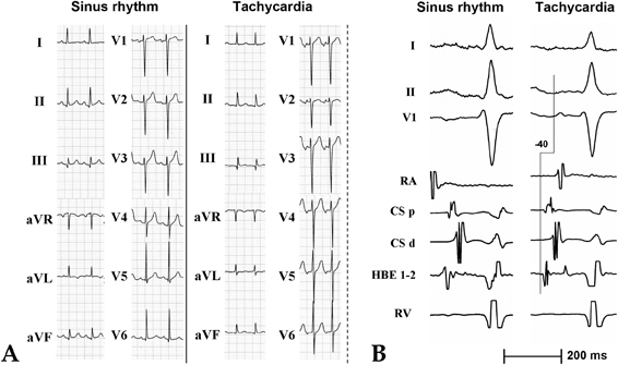
Fig. 1 (A) Twelve-lead electrocardiograms during sinus rhythm (Left) and the tachycardia (Right). (B) The tracings are the ECG leads I, II, V1, and intracardiac electrograms recorded from a mapping catheter, and catheters placed at the HBE, within the CS, and at the RVA during sinus rhythm (Left) and the tachycardia (Right). The local electrogram recorded at the His bundle preceded the onset of the surface P wave by 40 ms. ECG, electrocardiogram; CS, coronary sinus; HBE, His bundle region; RA, right atrium; RVA, right ventricular apex.
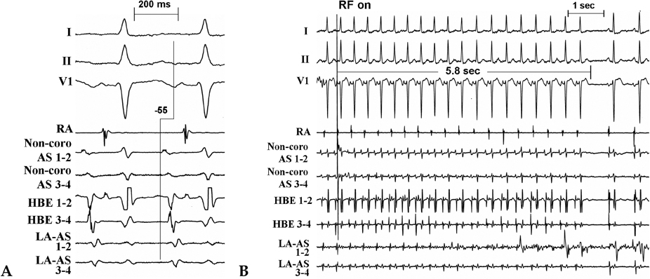
Fig. 2 The tracings are the ECG leads I, II, V1, and intracardiac electrograms recorded from a mapping catheter positioned in the non-coronary aortic sinus (Non-coro AS 1 to 2, Non-coro AS 3 to 4), and catheters placed at the HBE and left anteroseptum (LA AS 1 to 2, LA AS 3 to 4). (A) The successful site with the earliest RA activation is shown at the non-coronary aortic sinus. Note that the earliest activation preceded the onset of the surface P wave by 55 ms. (B) The atrial tachycardia was successfully terminated after 5.8 seconds during the first radiofrequency application. AS, aortic sinus; the other abbreviations are as in Fig. 1.
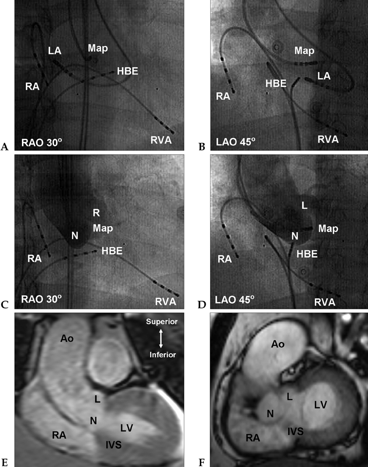
Fig. 3 Right (30°) and left (45°) oblique radiographic views showing the mapping catheter (Map) located at the successful ablation site in the aortic sinus (A and B), another mapping catheter at the left anteroseptum, and catheters placed at the HBE, in the RA, and in the RV. An aortic root angiogram in the right (30°) and left (45°) oblique radiographic views (C and D) showing the mapping catheter in the non-coronary aortic sinus supero-posterior to the site where the His bundle potential was recording from the distal one to four electrodes. An MRI in the right and left oblique views (E and F) showing the non-coronary aortic sinus. Ao, aorta; L, left aortic sinus; LV, left ventricle; N, non-coronary aortic sinus; R, right aortic sinus; IVS, interventricular septum; the other abbreviations are as in Fig. 1.
Cited by 1 articles
-
Prevalence and Characteristics of Atrial Tachycardia From Noncoronary Aortic Cusp During Atrial Fibrillation Catheter Ablation
Myung-Jin Cha, Jun Kim, Yoon Jung Park, Min Soo Cho, Hyoung-Seob Park, Soonil Kwon, Young Soo Lee, Jinhee Ahn, Hyung-Oh Choi, Jong-Sung Park, YouMi Hwang, Jin Hee Choi, Ki-Won Hwang, Yoo-Ri Kim, Seongwook Han, Seil Oh, Gi-Byoung Nam, Kee-Joon Choi, Hui-Nam Pak
Korean Circ J. 2022;52(7):513-526. doi: 10.4070/kcj.2021.0388.
Reference
-
1. Lai LP, Lin JL, Chen TF, Ko WC, Lien WP. Clinical, electrophysiological characteristics, and radiofrequency catheter ablation of atrial tachycardia near the apex of Koch's triangle. Pacing Clin Electrophysiol. 1998. 21:367–374.
Article2. Badhwar N, Kalman J, Sparks PB, Kistler PM, Attari M, Berger M, et al. Atrial tachycardia arising from the coronary sinus musculature: electrophysiological characteristics and long-term outcomes of radiofrequency ablation. J Am Coll Cardiol. 2005. 46:1921–1930.3. Connors SP, Vora A, Green MS, Tang AS. Radiofrequency ablation of atrial tachycardia originating from the triangle of Koch. Can J Cardiol. 2000. 16:39–43.4. Chen CC, Tai CT, Chiang CE, Yu WC, Lee SH, Chen YJ, et al. Atrial tachycardias originating from the atrial septum: electrophysiologic characteristics and radiofrequency ablation. J Cardiovasc Electrophysiol. 2000. 11:744–749.
Article5. Tada H, Naito S, Miyazaki A, Oshima S, Nogami A, Taniguchi K. Successful catheter ablation of atrial tachycardia originating near the atrioventricular node from the noncoronary sinus of Valsalva. Pacing Clin Electrophysiol. 2004. 27:1440–1443.
Article6. Ouyang F, Ma J, Ho SY, Bänsch D, Schmidt B, Ernst S, et al. Focal atrial tachycardia originating from the non-coronary arortic sinus, electrophysiological characteristics and catheter ablation. J Am Coll Cardiol. 2006. 48:122–131.
- Full Text Links
-
- Actions
-
Cited
- CITED
-
- Close
- Share
-
- Similar articles
-
- Atrial Tachycardia Originating from the Aortomitral Junction
- Radiofrequency Catheter Ablation of Atrial Tachycardia
- A case of sinus node dysfunction and atrial tachycardia after the excision of a left atrial myxoma
- Radiofrequency Catheter Ablation of Atrioventricular Nodal Reentrant Tachycardia in Two Patients with Persistent Left Superior Vena Cava
- Characteristics and Outcomes of Atrial Tachycardia Originating from the Sinus Venosus during Catheter Ablation of Atrial Fibrillation
