Cytologic Findings of Cervicovaginal Smears in Women with Uterine Papillary Serous Carcinoma
- Affiliations
-
- 1Department of Pathology, Samsung Cheil Hospital, Sungkyunkwan University School of Medicine, Seoul, Korea. ykcmd.chun@samsung.com
- KMID: 1781733
- DOI: http://doi.org/10.3346/jkms.2005.20.1.93
Abstract
- The goal of this study was to evaluate the cytomorphologic features of histologically confirmed uterine papillary serous carcinomas (UPSC) of the endometrium. We reviewed cervicovaginal smears from 12 patients with UPSC who had done their cervical smears at six months to a year earlier before the time of diagnosis; nine smears (75%) were diagnosed as positive for malignancy and three smears (25%) were diagnosed as negative. The cervical smears of patients with UPSC revealed frequent papillary clusters that were composed of large pleomorphic tumor cells with prominent nucleoli in a background of necrosis. Other findings revealed from the tests were relatively frequent single malignant cells and bare nuclei. Although the Pap smear is not a sensitive screening test for endometrial carcinoma, we could depend on it to reveal the cytologic features of UPSC which are fairly characteristic and reliable for a preoperative diagnosis of UPSC. Preoperative identification of this poor prognostic variant of endometrial carcinoma may influence the surgical management of these cases and the choice of adjuvant therapy.
MeSH Terms
-
Adenocarcinoma/diagnosis/pathology
Adult
Aged
Carcinoma
Carcinoma, Squamous Cell/diagnosis/pathology
Cystadenocarcinoma, Papillary/*diagnosis/*pathology
Cystadenocarcinoma, Serous/*diagnosis/*pathology
Diagnosis, Differential
Female
Humans
Middle Aged
Necrosis
Prognosis
Uterine Neoplasms/*diagnosis/*pathology
*Vaginal Smears
Figure
-

Fig. 1 Cervical smear of uterine papillary carcinoma shows papillary fragment of tumor cells with irregular outline and acute inflammatory cells in background (Papanicolaou stain, ×400).
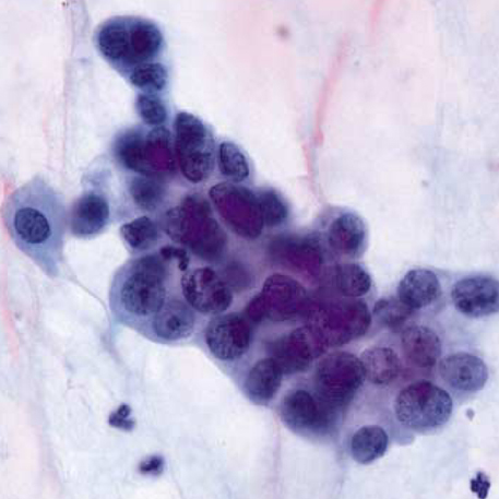
Fig. 2 Tumor cells of uterine papillary serous carcinoma have large nuclei with irregularly distributed chromatin, distinct nucleoli, irregular nuclear margins and indistinct cytoplasmic borders (Papanicolaou stain, ×1,000).

Fig. 3 Cervicovaginal smear of uterine papillary carcinoma shows a papillary cluster and single scattered malignant cells in background of tumor diathesis and lysed blood (Papanicolaou stain, ×400).

Fig. 4 Histology of uterine papillary serous carcinoma shows short and dense papillae. The cells covering the papillae and lining glands form small papillary tufts or floating clusters (H&E stain, ×100). Tumor cells reveal dense eosinophilic cytoplasm with high grade nulclear atypia (inset, H&E stain, ×400).
Reference
-
1. Christopherson WM, Alberhasky RC, Connelly PJ. Carcinoma of the endometrium. II. Papillary adenocarcinoma: a clinical pathological study, 46 cases. Am J Clin Pathol. 1982. 77:534–540.
Article2. Hendrickson M, Ross J, Eifel P, Martinez A, Kempson R. Uterine papillary serous carcinoma: a highly malignant form of endometrial adenocarcinoma. Am J Surg Pathol. 1982. 6:93–108.3. Walker AN, Mills SE. Serous papillary carcinoma of the endometrium. A clinicopathologic study of 11 cases. Diagn Gynecol Obstet. 1982. 4:261–267.4. Kuebler DL, Nikrui N, Bell DA. Cytologic features of endometrial papillary serous carcinoma. Acta Cytol. 1989. 33:120–126.5. Ronnett BM, Zaino RJ, Ellenson LH, Kurman RJ. Kurman RJ, editor. Endometrial carcinoma. Blaustein's Pathology of the Female Genital Tract. 2002. 5th ed. New York: Springer;528–533.6. Slomovitz BM, Burke TW, Eifel PJ, Ramondetta LM, Silva EG, Jhingran A, Oh JC, Atkinson EN, Broaddus RR, Gershenson DM, Lu KH. Uterine papillary serous carcinoma (UPSC): a single institution review of 129 cases. Gynecol Oncol. 2003. 91:463–469.
Article7. Lozowski MS, Mishriki Y, Solitare GB. Factors determining the degree of endometrial exfoliation and their diagnostic implications in endometrial adenocarcinoma. Acta Cytol. 1986. 30:623–627.8. Schneider ML, Wortmann M, Weigel A. Influence of the histologic and cytologic grade and the clinical and postsurgical stage on the rate of endometrial carcinoma detection by cervical cytology. Acta Cytol. 1986. 30:616–622.9. Gu M, Shi W, Barakat RR, Thaler HT, Saigo PE. Pap smears in women with endometrial carcinoma. Acta Cytol. 2001. 45:555–560.
Article10. Hong SR, Kim HS, Park JS. Significance of cytologic detection of endometrial carcinoma in papanicolaou smear: The relevance of histologic type, grade and stage. Korean J Cytopathol. 1993. 4:81–86.11. Zhou C, Matisic JP, Clement PB, Hayes MM. Cytologic features of papillary serous adenocarcinoma of the uterine cervix. Cancer. 1997. 81:98–104.
Article12. Wright CA, Leiman G, Burgess SM. The cytomorphology of papillary serous carcinoma of the endometrium in cervical smears. Cancer. 1999. 87:12–18.
Article13. Takashina T, Ono M, Kanda Y, Sagae S, Hayakawa O, Ito E. Cervicovaginal and endometrial cytology in ovarian cancer. Acta Cytol. 1988. 32:159–162.14. Sasagawa M, Nishino K, Honma S, Kodama S, Takahashi T. Origin of adenocarcinoma cells observed on cervical cytology. Acta Cytol. 2003. 47:410–414.
Article15. Takashina T, Ito E, Kudo R. Cytologic diagnosis of primary tubal cancer. Acta Cytol. 1985. 29:367–372.16. Hirai Y, Chen JT, Hamada T, Fujimoto I, Yamauchi K, Hasumi K, Masubuchi K, Sakamoto A. Clinical and cytologic aspects of primary fallopian tube carcinoma. A report of ten cases. Acta Cytol. 1987. 31:834–840.17. Seong SJ, Kim TJ, Lim KT, Chung HW, Lee KH, Park IS, Sihm JU, Park CT, Kim HS. Preoperative pap smears in endometrial carcinoma: The clinicopathologic relevance. Korean J Obstet Gynecol. 2002. 45:1746–1751.
