Korean J Lab Med.
2008 Oct;28(5):378-385. 10.3343/kjlm.2008.28.5.378.
Clinical and Cytogenetic Findings on 31,615 Mid-trimester Amniocenteses
- Affiliations
-
- 1Division of Cytogenetics, Department of Laboratory Medicine, Seoul Medical Science Institute, Seoul Clinical Laboratories, Seoul, Korea. dkrlee@scllab.co.kr
- 2Department of Laboratory Medicine, JungAng General Hospital, Jeju, Korea.
- KMID: 1781570
- DOI: http://doi.org/10.3343/kjlm.2008.28.5.378
Abstract
-
BACKGROUND: Since amniocentesis made prenatal diagnosis feasible in 1967, the method has become a popular tool in obstetric practices. In Korea, the demand for genetic counseling and prenatal tests has increased markedly because the number and proportion of pregnancies in women aged 35 yr and older have increased over a 20-yr period. Here we report clinical and cytogenetic findings on 31,615 mid-trimester amniocenteses.
METHODS
To investigate the changes in the annual number of amniocentesis, distribution of indications and age, and cytogenetic findings and abnormality rate according to indications, this study retrospectively analyzed 31,615 cases of mid-trimester amniocentesis performed at Seoul Clinical Laboratories, an independent medical laboratory center, during the past 13 yr (1994-2007).
RESULTS
The annual number of amniocenteses has increased substantially since 1994. Among the 31,615 amniocentesis cases, the maternal age between 30 and 34 yr was the most common age group (35.4%). Among clinical indications, abnormal maternal serum screening results have been the most common indication for amniocentesis since 1994. Chromosomal abnormalities were detected in 973 cases (3.1%). Down syndrome was the most common abnormality found (36.9%, 359/973). In sex chromosomal abnormalities, 53 cases of Turner syndromes, 32 cases of Klinefelter syndromes, 20 cases of triple X syndromes, and 15 cases of 47,XYY were diagnosed. Of structural rearrangements, reciprocal translocations between two autosomes were the most common (15.5%, 151/973). Abnormal ltrasonographic findings showed the highest positive predictive value (5.9%) among the clinical indications.
CONCLUSIONS
The present study could be used for the establishment of a database for genetic counseling. The discovery of an abnormality provides the option of termination or continuation in the pregnancy, a more suitable obstetric management in Korea.
MeSH Terms
Figure
-
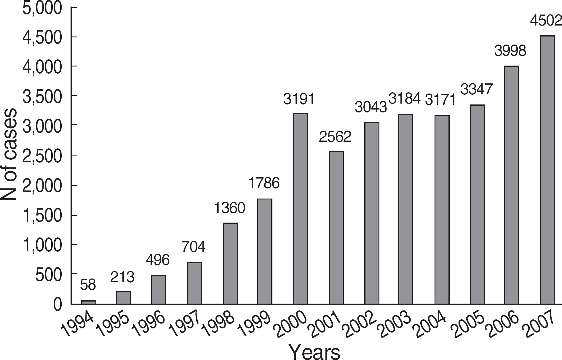
Fig. 1. Annual distribution of amniocentesis cases in Seoul Clinical Laboratories, Seoul, Korea.
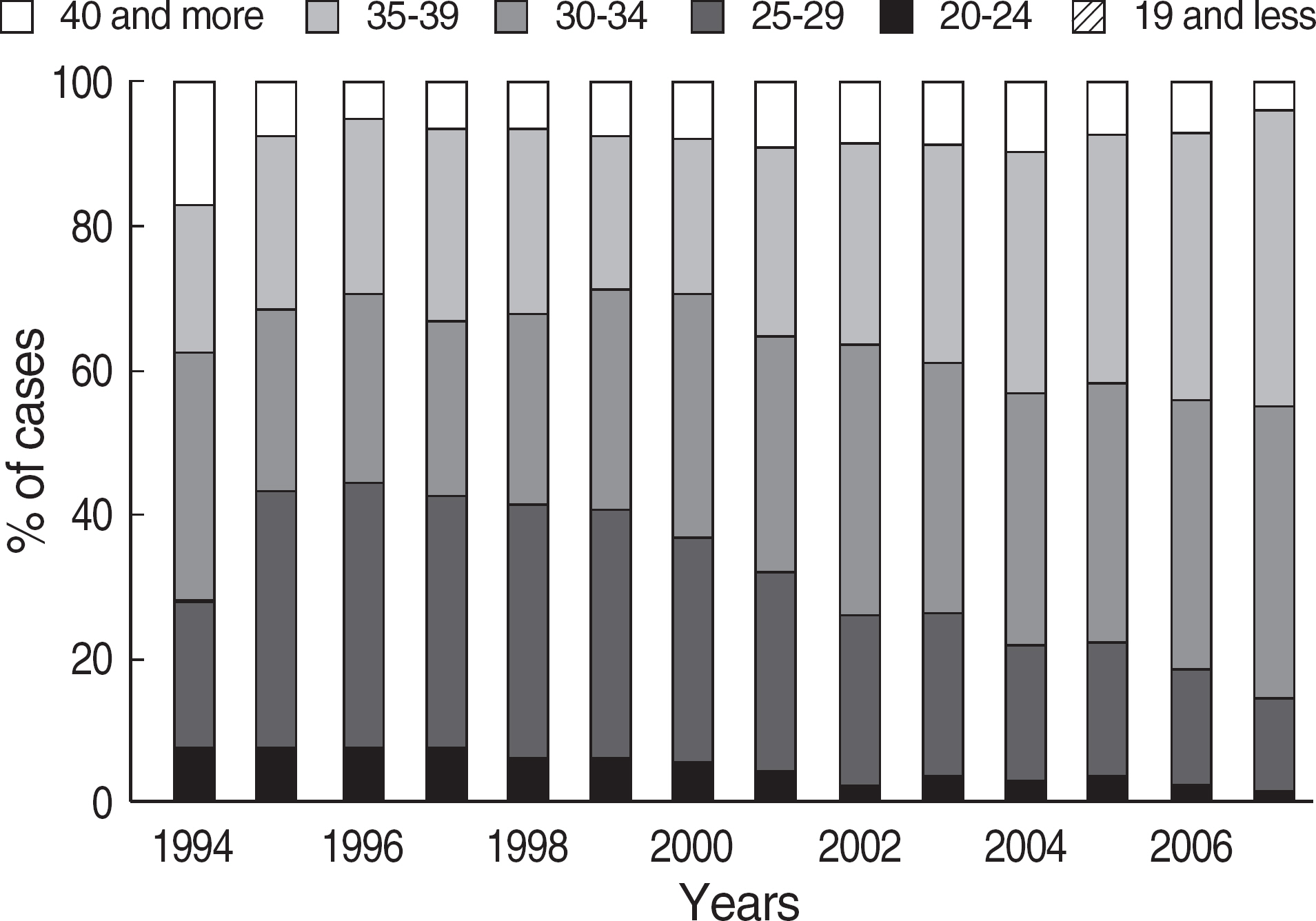
Fig. 2. Annual distribution of maternal age group.
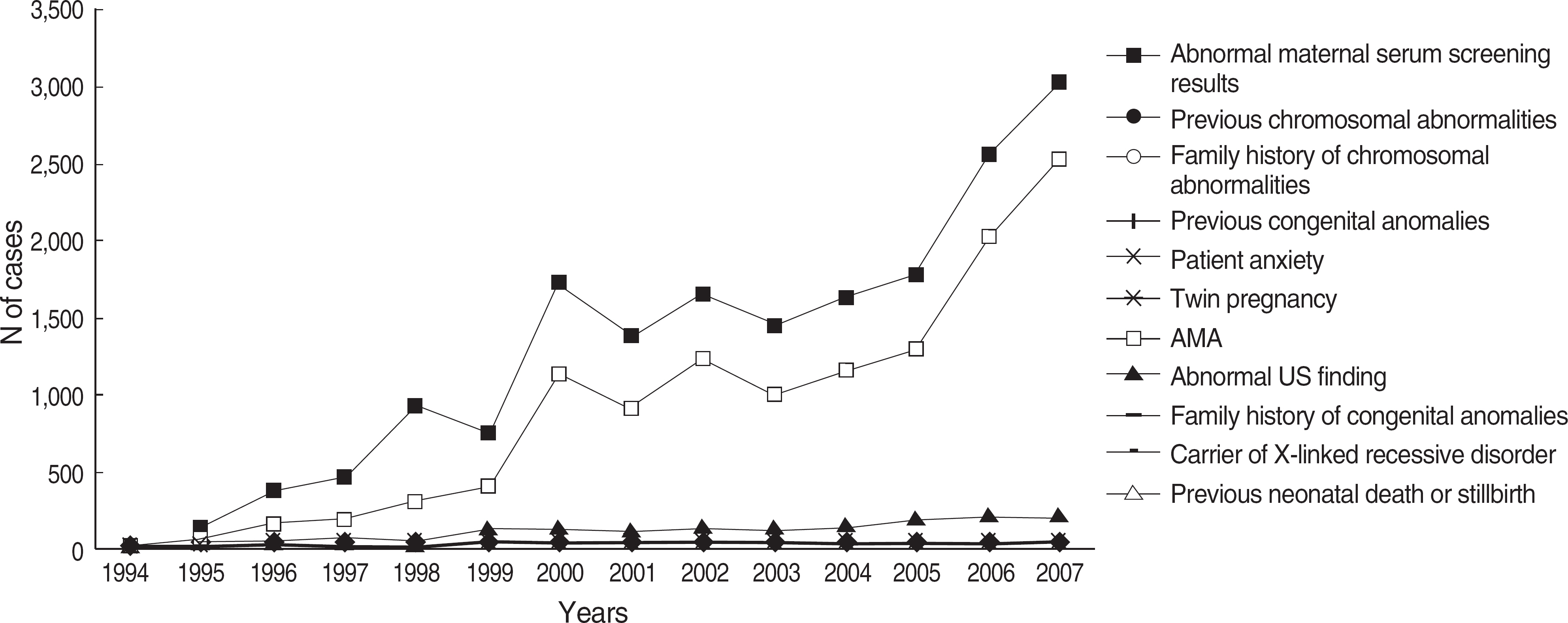
Fig. 3. Annual distribution of amniocentesis cases by clinical indications.
Cited by 1 articles
-
Performance Characteristics of the UniCel DxI 800 Immunoassay for the Maternal Serum Quadruple Test, Including Median Values for Each Week of Gestation, in Korean Women
Jeong Hoon Lee, Yongjung Park, Borum Suh, Seon-Mi Song, Oh Hun Kwon, Jeong-Ho Kim
Korean J Lab Med. 2010;30(2):126-132. doi: 10.3343/kjlm.2010.30.2.126.
Reference
-
1.Dallaire L. Integration of prenatal diagnosis of genetic diseases into medical practice. Can Med Assoc J. 1976. 115:713–4.2.Caron L., Tihy F., Dallaire L. Frequencies of chromosomal abnormalities at amniocentesis: over 20 years of cytogenetic analyses in one laboratory. Am J Med Genet. 1999. 15:149–54.
Article3.Hook EB., Hamerton JL. The frequency of chromosome abnormalities detected in consecutive newborn studies, difference between studies results by sex and severity of phenotypic involvement. Hook EB, Porter H, editors. Population cytogenetics studies in humans. 1st ed.New York: Academic Press;1977. p. 63–79.4.Hook EB., Cross PK., Schreinemachers DM. Chromosomal abnormality rates at amniocentesis and in live-born infants. JAMA. 1983. 249:2034–8.
Article5.Milunsky A., Atkins L. The frequency of chromosomal abnormalities diagnosed prenatally. Hook EB, Porter H, editors. Population cytogenetics, studies in humans. 1st ed.New York: Academic Press;1977. p. 11–25.6.Verma RS., Babu A. Tissue culture techniques and chromosome preparation. Verma RS, Babu A, editors. Human chromosomes principles and techniques. 2nd ed.New York: McGraw-Hill Press;1995. p. 6–71.7.Seabright M. A rapid banding technique for human chromosomes. Lancet. 1971. 30:971–2.
Article8.Karaoguz MY., Bal F., Yakut T., Ercelen NO., Ergun MA., Gokcen AB, et al. Cytogenetic results of amniocentesis materials: incidence of abnormal karyotypes in the Turkish collaborative study. Genet Couns. 2006. 17:219–30.9.Tseng JJ., Chou MM., Lo FC., Lai HY., Chen MH., Ho ES. Detection of chromosome aberrations in the second trimester using genetic amniocentesis: experience during 1995-2004. Taiwan J Obstet Gynecol. 2006. 45:39–41.
Article10.Jang SK., Choi OH. Cytogenetic and clinical analysis in 3,537 cases of midtrimester amniocentesis. Korean J Perinatol. 2007. 18:29–36. (장성규 및 최욱환. 임신 중기 양수천자 3,537예에 대한 세포유전학적분석및임상적고찰. 대한주산의학회잡지 2007;18: 29-36.).11.Choi SJ., Kim WS., Kim JU., Lee ES., Cho EH., Kim SH, et al. Cytogenetic and clinical analysis of midtrimester amniocentesis. Korean J Obstet Gynecol. 2005. 48:1420–30. (최석주, 김우선, 김지운, 이은실, 조은해, 김선희 등. 임신 중기 양수천자의 세포유전학적 분석 및 임상적 고찰. 대한산부인과학회지 2005;48: 1420-30.).12.Shim JY., Kim SH., Kim JS., Ahn SM., Seo EJ., Yoo HW, et al. Clinical analysis of prenatal cytogenetic diagnoses: four-year experience at Asan medical center. Korean J Obstet Gynecol. 2004. 47:487–94. (심재윤, 김성훈, 김정숙, 안송미, 서을주, 유한욱 등. 산전 세포유전학적 검사의 임상적 분석: 서울아산병원 4년간의 경험. 대한산부인과학회지 2004;47: 487-94.).13.Park IY., Shin JC., Kim SC., Ahn HY., Moon HB., Park CH, et al. Cytogenetic analysis in 3,503 cases of midtrimester amniocentesis: CUMC experience (II). Korean J Obstet Gynecol. 2004. 47:96–103. (박인양, 신종철, 김석찬, 안현영, 문희봉, 박철훈 등. 임신 중기 양수천자 3,503예에대한 세포 유전학적 분석: CUMC 경험(II). 대한산부인과학회지 2004;47: 96-103.).14.Kim YJ. Cytogenetic analysis in 651 cases of amniocentesis. Korean J Lab Med. 2002. 22:208–12. (김영재. 양수천자 651예의 세포유전학적분석. 대한진단검사의학회지 2002;22: 208-12.).15.Kim HJ., Lee SM., Kim EJ., Cho EN., Park SY., Kang KH, et al. A review of prenatal cytogenetic analysis in 2942 midtrimester amniocentesis. Korean J Obstet Gynecol. 2001. 44:1109–14. (김현진, 이승무, 김은정, 조은나, 박소양, 강경화등. 임신중기양수천자 2942예의세포유전학적연구. 대한산부인과학회지 2001;44: 1109-14.).16.Midtrimester amniocentesis for prenatal diagnosis. Safety and accuracy. JAMA. 1976. 236:1471–6.17.Simpson NE., Dallaire L., Miller JR., Siminovich L., Hamerton JL., Miller J, et al. Prenatal diagnosis of genetic disease in Canada: report of a collaborative study. Can Med Assoc J. 1976. 23:739–48.18.Dewald G. Cytogenetics. McClatchey KD, editor. Clinical laboratory medicine. 1st ed.Baltimore: Williams & Wilkins;1994. p. 643–5.19.Gardner RJM, Sutherland GR, editors. Chromosome abnormalities and genetic counseling. 3rd ed.New York: Oxford University Press;2004. p. 252–6.20.Unger E., Pincus MR. Molecular pathology. Mcpherson RA, Pincus MR, editors. Clinical diagnosis and management by laboratory methods. 21st ed.Philadelphia: WB Saunders;2007. p. 1276–7.21.Jacobs P., Dalton P., James R., Mosse K., Power M., Robinson D, et al. Turner syndrome: a cytogenetic and molecular study. Ann Hum Genet. 1997. 6:471–83.
Article22.Sybert VP. Turner syndrome. Cassidy SB, Allanson JE, editors. Management of genetic syndromes. 1st ed.New York: Wiley-Liss;2001. p. 459–84.
Article23.Held KR., Kerber S., Kaminsky E., Singh S., Goetz P., Seemanova E, et al. Mosaicism in 45,X Turner syndrome: does survival in early pregnancy depend on the presence of two sex chromosomes? Hum Genet. 1992. 88:288–94.
Article24.Fernandez-Garcia R., Garcia-Doval S., Costoya S., Pasaro E. Analysis of sex chromosome aneuploidy in 41 patients with Turner syndrome: a study of ‘hidden’ mosaicism. Clin Genet. 2000. 58:201–8.25.Baena N., De Vigan C., Cariati E., Clementi M., Stoll C., Caballin MR, et al. Turner syndrome: evaluation of prenatal diagnosis in 19 European registries. Am J Med Genet A. 2004. 129:16–20.26.Gravholt CH., Fedder J., Naeraa RW., Muller J. Occurrence of gona-doblastoma in females with Turner syndrome and Y chromosome material: a population study. J Clin Endorinol Metab. 2000. 85:3199–202.
Article27.Huang B., Thangavelu M., Bhatt S., J Sandlin C., Wang S. Prenatal diagnosis of 45,X and 45,X mosaicism: the need for thorough cytogenetic and clinical evaluations. Prenata Diagn. 2002. 22:105–10.
Article28.Hsu LY. Prenatal diagnosis of chromosomal abnormalities through amniocentesis. Milunsky A, editor. Genetic disorders and the fetus: diagnosis, prevention, and treatment. 4th ed.Baltimore: The Johns Hopkins University Press;1998. p. 179–248.29.Crolla JA. FISH and molecular studies of autosomal supernumerary marker chromosomes excluding those derived from chromosome 15: II. Review of the literature. Am J Med Genet. 1998. 75:367–81.
Article30.Huang B., Solomon S., Thangavelu M., Peters K., Bhatt S. Supernumerary marker chromosomes detected in 100,000 prenatal diagnoses: molecular cytogenetic studies and clinical significance. Prenata Diagn. 2006. 26:1142–50.31.Yang YH., Ju KS., Kim SB., Cho YH., Lee JH., Lee SH, et al. The Korean collaborative study on 11,000 prenatal genetic amniocentesis. Yonsei Med J. 1999. 40:460–6.
Article
- Full Text Links
-
- Actions
-
Cited
- CITED
-
- Close
- Share
-
- Similar articles
-
- Comparison of Early and Mid-Second-Trimester Amniocentesis
- Prenatal screening for neural tube defects: from maternal serum alpha-fetoprotein to ultrasonography
- The differences of mid-trimester triple marker levels according to the occurrence time and severity of preeclampsia
- A Case of Idiopathic Acute Hepatitis with Complications in Mid-Trimester Pregnancy
- Six-years' Experience of Pseudomosaicism and Maternal Cell Contamination in Cultured Amniocytes
