Prenatal screening for neural tube defects: from maternal serum alpha-fetoprotein to ultrasonography
- Affiliations
-
- 1Department of Obstetrics and Gynecology, Chung-Ang University College of Medicine, Seoul, Korea
- 2Chung-Ang University Gwangmyeong Hospital, Gwangmyeong, Korea
- KMID: 2538343
- DOI: http://doi.org/10.5468/ogs.22263
Abstract
- The two main screening tests during pregnancy are those for chromosomal abnormalities and neural tube defects (NTDs). In particular, for NTDs, measurement of maternal serum alpha-fetoprotein (MSAFP) levels early in the second trimester (15-18 weeks of gestation) has been considered the gold standard screening test for the past 4 decades. However, with remarkable technological advancements and the widespread use of ultrasound during those periods, mid-trimester ultrasonography has gradually replaced the role of measuring MSAFP levels as a screening method for NTDs. This change was initiated more about 10 years ago in some countries, which have issued national guidelines to use mid-trimester ultrasonography instead of measuring MSAFP levels as a prenatal screening method for NTDs. However, no significant changes have occurred in Korea, where second-trimester ultrasonography is routinely performed with high-quality equipment. We aimed to provide information regarding the importance of changing the screening method for NTDs from MSAFP measurement to ultrasonography, and to detail methods of implementing mid-trimester ultrasonography for screening purposes. We also share our experience of operating a prenatal diagnostic program for NTDs without using MSAFP for more than 15 years.
Figure
-
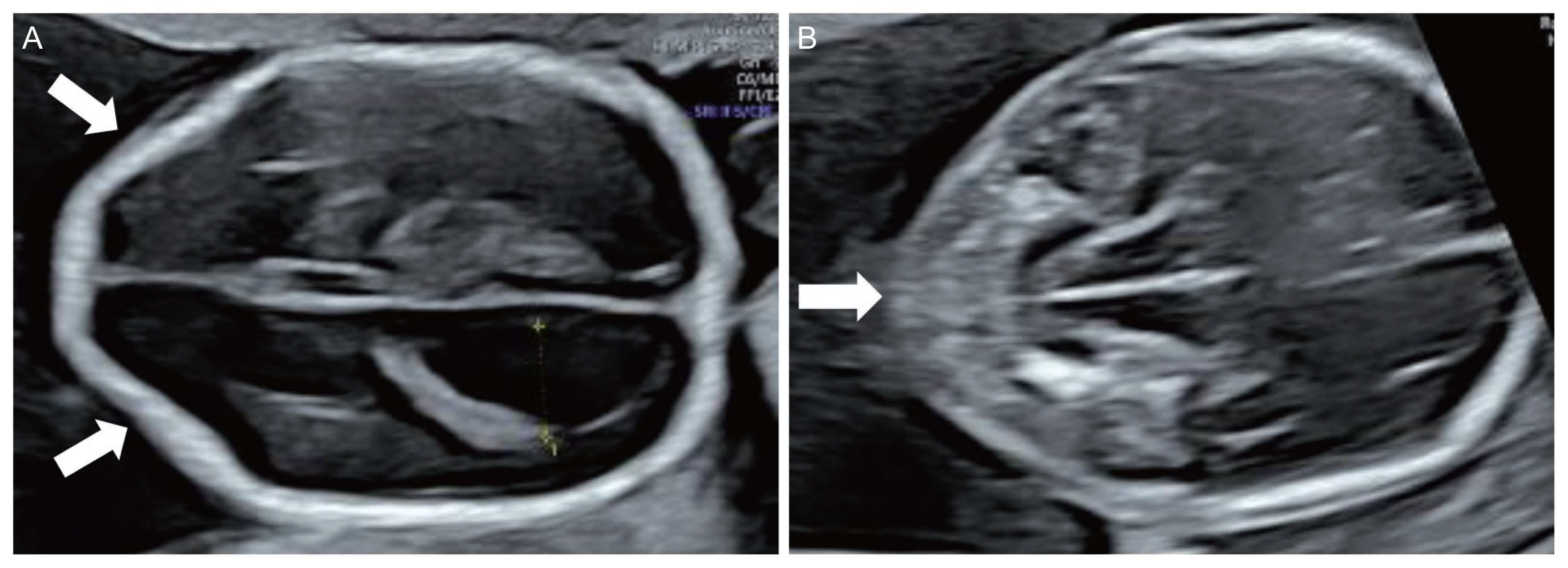
Fig. 1 Indirect brain signs of the fetus affected by open spinal neural tube defects. (A) Abnormal skull shape (lemon sign, arrows) and ventriculomegaly at 18 gestational weeks; (B) abnormal cerebellum (banana sign, arrow) at 19 gestational weeks.
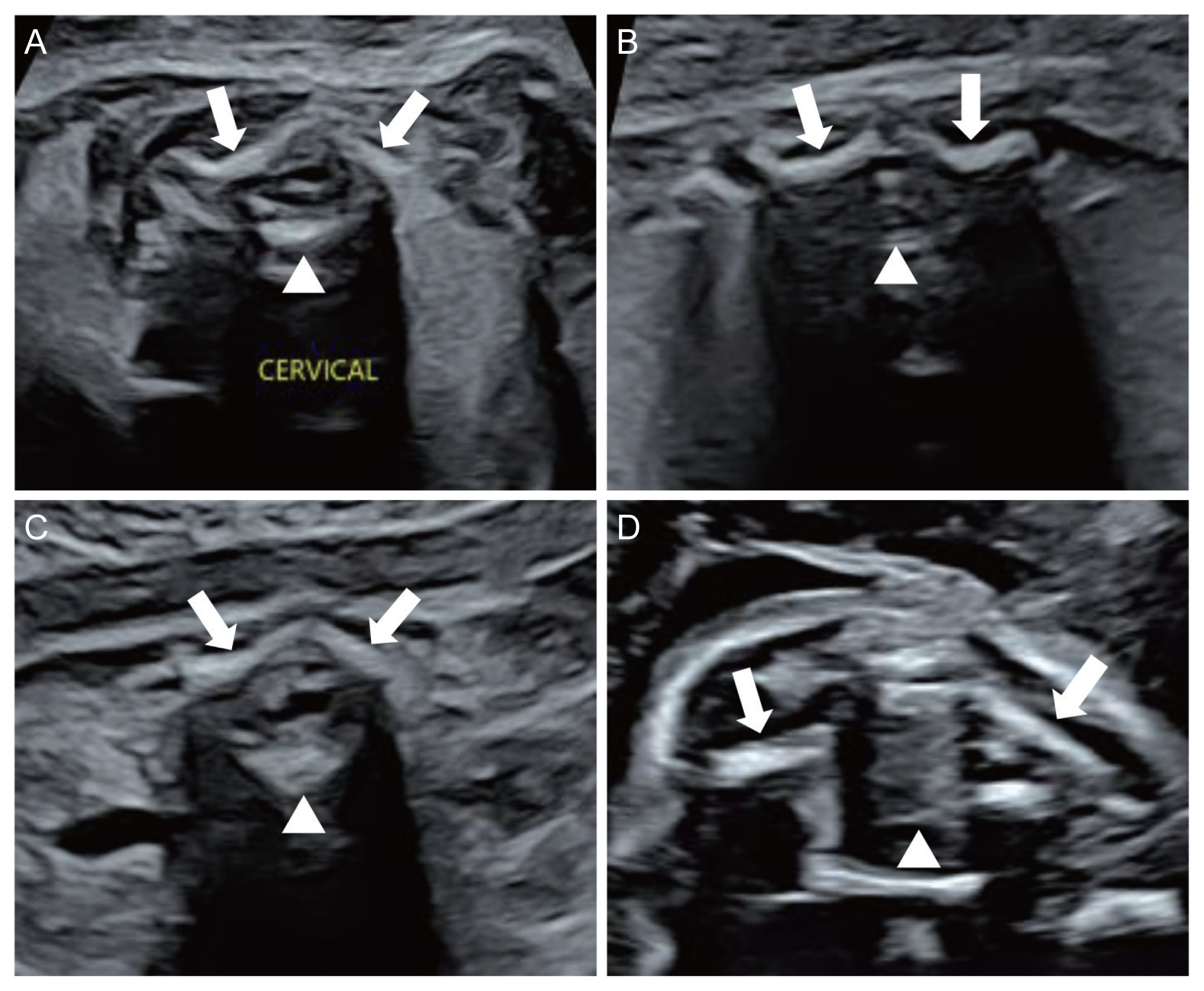
Fig. 2 Axial planes of a normal fetal spine at 25 gestational weeks. (A) Axial plane view of the cervical spine; (B) axial plane view of the thoracic spine; (C) axial plane view of the lumbar spine; (D) axial plane view of the sacrum (arrows: pedicles, arrowhead: vertebral body).
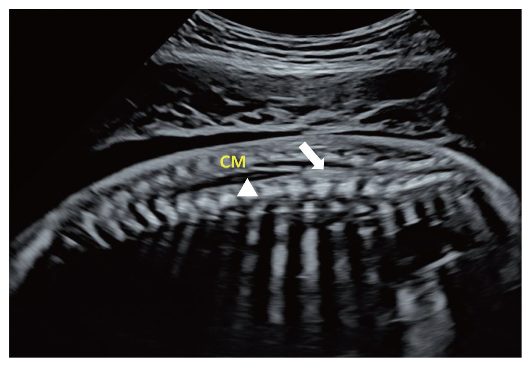
Fig. 3 Sagittal plane view of the normal spine and conus medullaris (CM) at 22 gestational weeks (arrow: filum terminale, arrowhead: central canal).
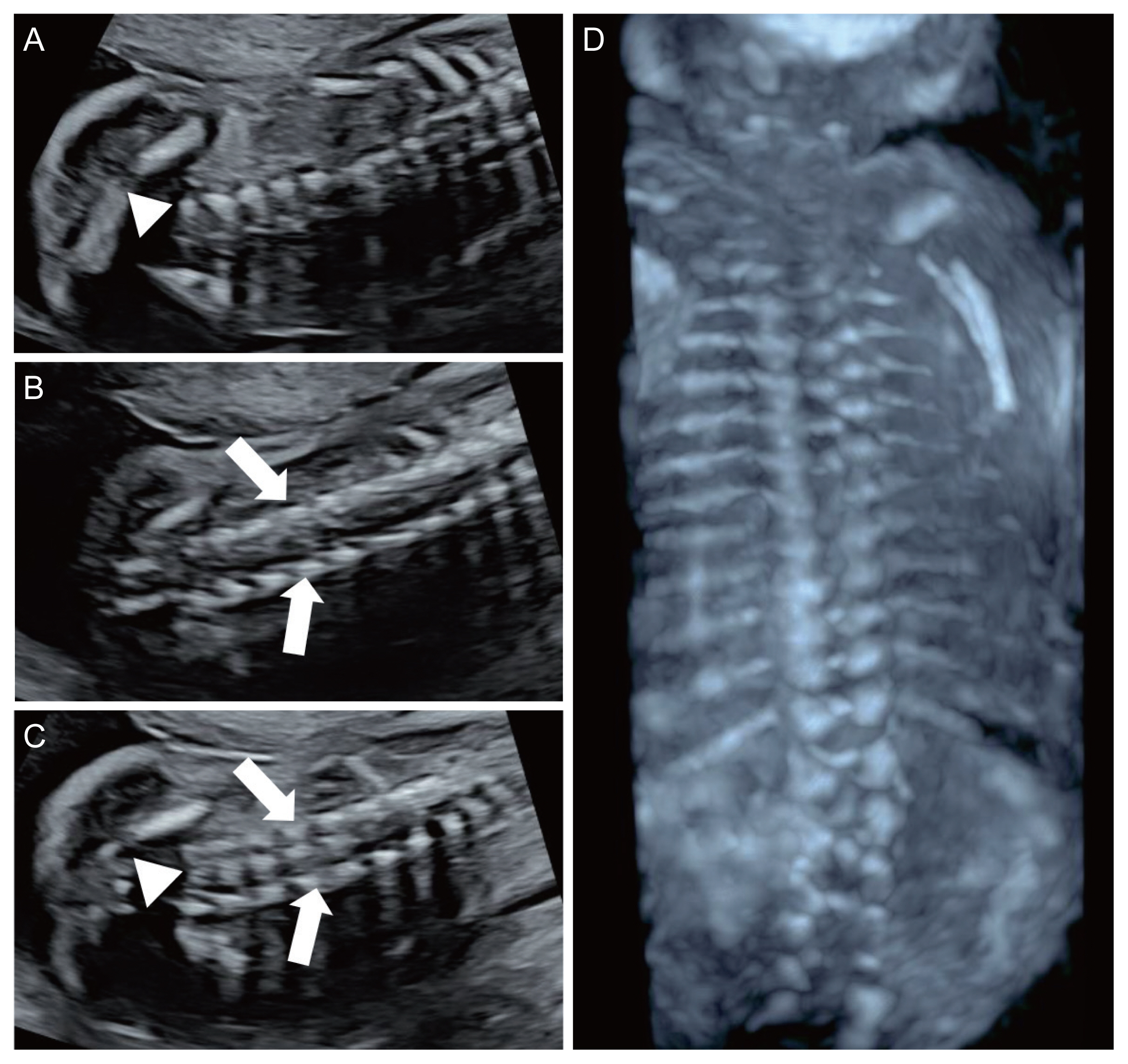
Fig. 4 Coronal plane views of the lumbosacral spine at 25 gestational weeks. (A) Single line (arrowhead: vertebral body) of ossification; (B) double line (arrows: pedicles) of ossification; (C) triple line of ossification (arrows: pedicles, arrowhead: vertebral body). (D) Three-dimensional image of the fetal spine at 21 gestational weeks.
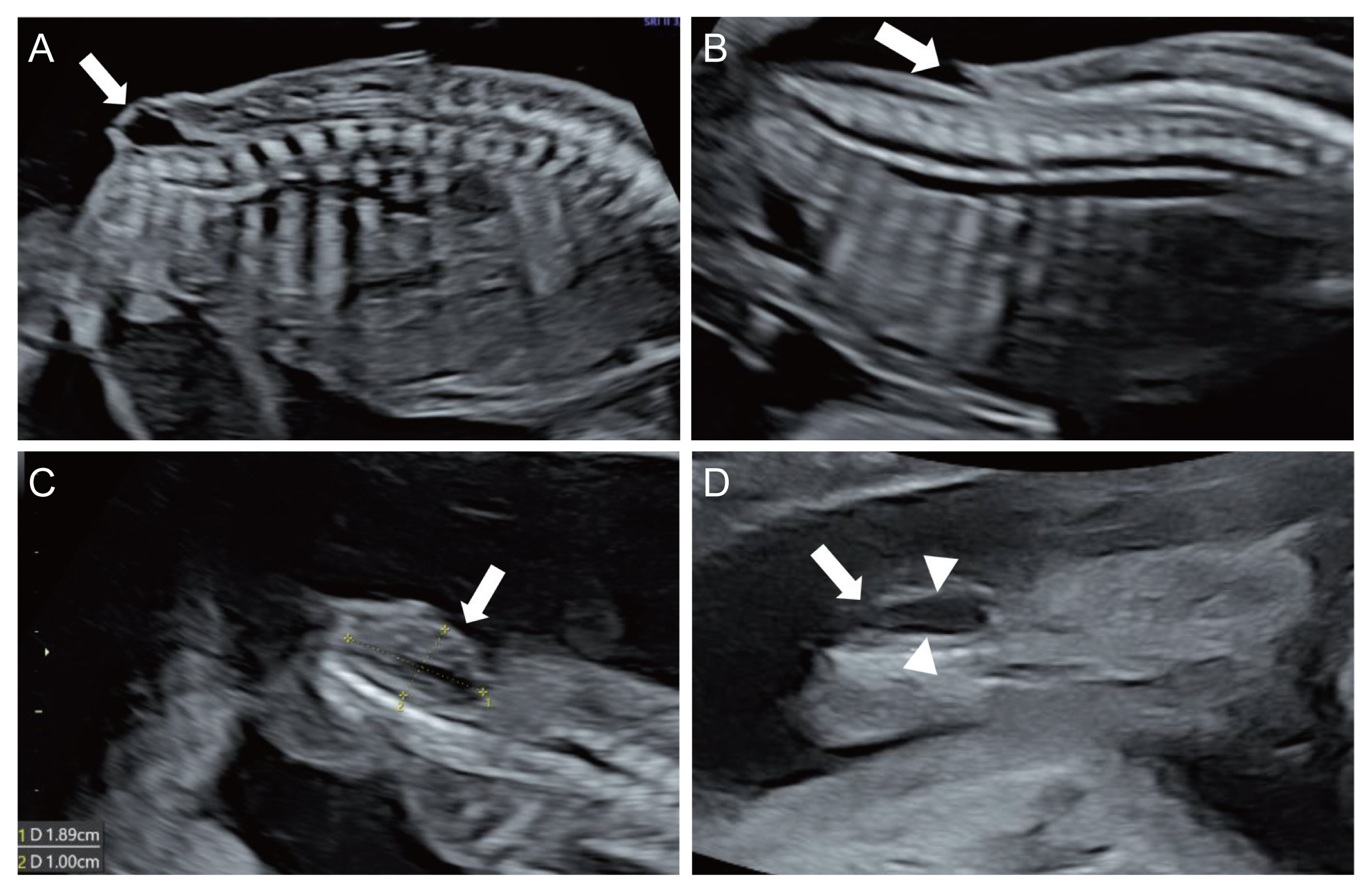
Fig. 5 Ultrasound images of spinal neural tube defects. (A) Protruding cystic mass (arrow) in sagittal plane; (B) indented skin line (arrow) in the sagittal plane; (C) indented skin (arrow) in the coronal plane; (D) ruptured meningomyelocele (arrow) with contour of the lesion (arowheads) in the coronal plane.
Reference
-
References
1. Driscoll DA, Gross SJ. Screening for fetal aneuploidy and neural tube defects. Genet Med. 2009; 11:818–21.
Article2. Practice bulletin No. 187: neural tube defects. Obstet Gynecol. 2017; 130:e279–90.3. Cameron M, Moran P. Prenatal screening and diagnosis of neural tube defects. Prenat Diagn. 2009; 29:402–11.
Article4. Wilson RD. Prenatal screening, diagnosis, and pregnancy management of fetal neural tube defects. J Obstet Gynaecol Can. 2014; 36:927–39.5. Cheschier N. ACOG practice bulletin. Neural tube defects. Int J Gynaeco Obstet. 2003; 83:123–33.6. Norem CT, Schoen EJ, Walton DL, Krieger RC, O’Keefe J, To TT, et al. Routine ultrasonography compared with maternal serum alpha-fetoprotein for neural tube defect screening. Obstet Gynecol. 2005; 106:747–52.
Article7. Yagel S, Valsky DV. ISUOG practice guidelines (updated): sonographic examination of the fetal central nervous system. Part 1: performance of screening examination and indications for targeted neurosonography. Ultrasound Obstet Gynecol. 2021; 57:173–4.8. Paladini D, Malinger G, Birnbaum R, Monteagudo A, Pilu G, Salomon LJ, et al. ISUOG practice guidelines (updated): sonographic examination of the fetal central nervous system. Part 2: performance of targeted neurosonography. Ultrasound Obstet Gynecol. 2021; 57:661–71.9. Douglas Wilson R, Van Mieghem T, Langlois S, Church P. Guideline no. 410: prevention, screening, diagnosis, and pregnancy management for fetal neural tube defects. J Obstet Gynaecol Can. 2021; 43:124–39.
Article10. Habib ZA. Maternal serum alpha-feto-protein: its value in antenatal diagnosis of genetic disease and in obstetrical-gynaecological care. Acta Obstet Gynecol Scand Suppl. 1977; 61:1–92.
Article11. Bergstrand CG, Czar B. Demonstration of a new protein fraction in serum from the human fetus. Scand J Clin Lab Invest. 1956; 8:174.
Article12. Gitlin D, Boesman M. Serum alpha-fetoprotein, albumin, and gamma-G-globulin in the human conceptus. J Clin Invest. 1966; 45:1826–38.
Article13. Johansson SG, Kjessler B, Sherman MS, Wahlström J. Alpha-fetoprotein (AFP) levels in maternal serum and amniotic fluid in singleton pregnant women in their 10th-25th week post last menstrual period. Acta Obstet Gynecol Scand Suppl. 1977; 69:20–4.
Article14. Kim CK, Yang YH. Alpha-fetoprotein values in maternal serum and amniotic fluid for prenatal screening of genetic disorders. Yonsei Med J. 1987; 28:218–27.
Article15. Leighton PC, Kitau MJ, Chard T, Gordon YB, Leek AE. Levels of alpha-fetoprotein in maternal blood as a screening test for fetal neural-tube defect. Lancet. 1975; 2:1012–5.
Article16. Wald NJ, Cuckle H, Brock JH, Peto R, Polani PE, Woodford FP. Maternal serum-alpha-fetoprotein measurement in antenatal screening for anencephaly and spina bifida in early pregnancy. Report of U.K. collaborative study on alpha-fetoprotein in relation to neural-tube defects. Lancet. 1977; 1:1323–32.17. Bradley LA, Palomaki GE, McDowell GA. Technical standards and guidelines: prenatal screening for open neural tube defects. Genet Med. 2005; 7:355–69.18. Wald N, Cuckle H, Boreham J, Stirrat G. Small biparietal diameter of fetuses with spina bifida: implications for antenatal screening. Br J Obstet Gynaecol. 1980; 87:219–21.
Article19. Nicolaides KH, Campbell S, Gabbe SG, Guidetti R. Ultrasound screening for spina bifida: cranial and cerebellar signs. Lancet. 1986; 2:72–4.
Article20. Warder DE. Tethered cord syndrome and occult spinal dysraphism. Neurosurg Focus. 2001; 10:e1.
Article21. Richards DS, Seeds JW, Katz VL, Lingley LH, Albright SG, Cefalo RC. Elevated maternal serum alpha-fetoprotein with normal ultrasound: is amniocentesis always appropriate? A review of 26,069 screened patients. Obstet Gynecol. 1988; 71:203–7.22. Puntachai P, Wanapirak C, Sirichotiyakul S, Tongprasert F, Srisupundit K, Luewan S, et al. Associations between pregnancy outcomes and unexplained high and low maternal serum alpha-fetoprotein levels. Arch Gynecol Obstet. 2015; 292:81–5.
Article23. Gagnon A, Wilson RD. Obstetrical complications associated with abnormal maternal serum markers analytes. J Obstet Gynaecol Can. 2008; 30:918–32.24. Chandra S, Scott H, Dodds L, Watts C, Blight C, Van den Hof M. Unexplained elevated maternal serum alphafetoprotein and/or human chorionic gonadotropin and the risk of adverse outcomes. Am J Obstet Gynecol. 2003; 189:775–81.25. Katz VL, Chescheir NC, Cefalo RC. Unexplained elevations of maternal serum alpha-fetoprotein. Obstet Gynecol Surv. 1990; 45:719–26.
Article26. Hogge WA, Thiagarajah S, Ferguson JE 2nd, Schnatterly PT, Harbert GM Jr. The role of ultrasonography and amniocentesis in the evaluation of pregnancies at risk for neural tube defects. Am J Obstet Gynecol. 1989; 161:520–3.
Article27. Crane JP, LeFevre ML, Winborn RC, Evans JK, Ewigman BG, Bain RP, et al. A randomized trial of prenatal ultrasonographic screening: impact on the detection, management, and outcome of anomalous fetuses. The RADIUS study group. Am J Obstet Gynecol. 1994; 171:392–9.28. Grandjean H, Larroque D, Levi S. The performance of routine ultrasonographic screening of pregnancies in the Eurofetus study. Am J Obstet Gynecol. 1999; 181:446–54.
Article29. Lennon CA, Gray DL. Sensitivity and specificity of ultrasound for the detection of neural tube and ventral wall defects in a high-risk population. Obstet Gynecol. 1999; 94:562–6.
Article30. Dashe JS, Twickler DM, Santos-Ramos R, McIntire DD, Ramus RM. Alpha-fetoprotein detection of neural tube defects and the impact of standard ultrasound. Am J Obstet Gynecol. 2006; 195:1623–8.
Article31. Arnautovic A, Splavski B, Boop FA, Arnautovic KI. Pediatric and adult chiari malformation type I surgical series 1965–2013: a review of demographics, operative treatment, and outcomes. J Neurosurg Pediatr. 2015; 15:161–77.
Article32. Langridge B, Phillips E, Choi D. Chiari malformation type 1: a systematic review of natural history and conservative management. World Neurosurg. 2017; 104:213–19.
Article33. McLone DG, Knepper PA. The cause of Chiari II malformation: a unified theory. Pediatr Neurosci. 1989; 15:1–12.
Article34. D’Addario V, Rossi AC, Pinto V, Pintucci A, Di Cagno L. Comparison of six sonographic signs in the prenatal diagnosis of spina bifida. J Perinat Med. 2008; 36:330–4.35. Kim GJ, Lee ES, Kim DH, Lee SH, Park JD, Park MH. Factors related with intracranial signs in fetuses with open neural tube defect. Obstet Gynecol Sci. 2005; 48:2541–9.36. Kollias SS, Goldstein RB, Cogen PH, Filly RA. Prenatally detected myelomeningoceles: sonographic accuracy in estimation of the spinal level. Radiology. 1992; 185:109–12.37. Stephenson SR. Sonographic signs of fetal neural tube and central nervous system defects. J Diagn Med Sonogr. 2003; 19:347–57.
Article38. Coleman BG, Langer JE, Horii SC. The diagnostic features of spina bifida: the role of ultrasound. Fetal Diagn Ther. 2015; 37:179–96.
Article39. Fong KW, Toi A, Okun N, Al-Shami E, Menezes RJ. Retrospective review of diagnostic performance of intracranial translucency in detection of open spina bifida at the 11–13-week scan. Ultrasound Obstet Gynecol. 2011; 38:630–4.
Article40. Mangione R, Dhombres F, Lelong N, Amat S, Atoub F, Friszer S, et al. Screening for fetal spina bifida at the 11–13-week scan using three anatomical features of the posterior brain. Ultrasound Obstet Gynecol. 2013; 42:416–20.41. Kappou D, Papastefanou I, Pilalis A, Kavalakis I, Kassanos D, Souka AP. Towards detecting open spina bifida in the first trimester: the examination of the posterior brain. Fetal Diagn Ther. 2015; 37:294–300.
Article42. Williams MA, Hickok DE, Zingheim RW, Luthy DA, Kimelman J, Nyberg DA, et al. Elevated maternal serum alpha-fetoprotein levels and midtrimester placental abnormalities in relation to subsequent adverse pregnancy outcomes. Am J Obstet Gynecol. 1992; 167:1032–7.
- Full Text Links
-
- Actions
-
Cited
- CITED
-
- Close
- Share
-
- Similar articles
-
- A Case of Spina Bifida Diagnosed by Prenatal Ultrasonography
- A meningomyelocele with normal intracranial signs on ultrasound and false-negative amniotic fluid alpha-fetoprotein and acetylcholinesterase
- A case of false positive amniotic acetylcholinesterase in a normal pregnancy
- Alpha-Fetoprotein Values in Maternal Serum and Amniotic Fluid for Prenatal Screening of Genetic Disorders
- A Case of Iniencephaly Diagnosed by Prenatal Ultrasonography
