Modifications of T-Scores by Quantitative Ultrasonography for the Diagnosis of Osteoporosis in Koreans
- Affiliations
-
- 1Department of Internal Medicine, College of Medicine, Yonsei University, Seoul, Korea. lsk@yuhs.ac
- 2Endocrine Research Institute, Brain Korea 21 Project for Medical Science, College of Medicine, Yonsei University, Seoul, Korea.
- 3Division of Endocrinology, Department of Internal Medicine, Dongeui Medical Center, Busan, Korea.
- 4Department of Internal Medicine, Cheil General Hospital & Women's Healthcare Center, Kwandong University College of Medicine, Seoul, Korea.
- 5Division of Endocrinology, Department of Internal Medicine, National Health Insurance Corporation Ilsan Hospital, Goyang, Korea.
- KMID: 1779123
- DOI: http://doi.org/10.3346/jkms.2009.24.2.232
Abstract
- To identify a proper T-score threshold for the diagnosis of osteoporosis in Koreans using quantitative ultrasonography (QUS), normative data from 240 females and 238 males (ages 20-29 yr) were newly generated. Then, the osteoporosis prevalence estimate for men and women over 50 yr of age was analyzed using previous World Health Organization (WHO) methods and heel QUS. T-scores were calculated from the normative data. There were definite negative correlations between age and all of the QUS parameters, such as speed of sound (SOS), broadband ultrasound attenuation (BUA), and estimated heel bone mineral density (BMD) (p<0.0001). After applying the recently determined prevalence of incident vertebral fracture in Koreans over 50 yr of age (11.6% and 9.1%, female vs male, respectively) to the diagnosis of osteoporosis by T-scores from heel BMD as measured by QUS, it was revealed that applicable T-scores for women and men were -2.25 and -1.85, respectively. These data suggest that simply using a T-score of -2.5, the classical WHO threshold for osteoporosis, underestimates the true prevalence when using peripheral QUS. Further prospective study of the power of QUS in predicting the absolute risk of fracture is needed.
Keyword
MeSH Terms
Figure
-

Fig. 1 Normal distribution curve of the QUS parameters in the female reference group. (A) Estimated heel BMD, (B) Speed of sound, (C) Broadband ultrasound attenuation.
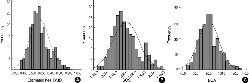
Fig. 2 Normal distribution curve of the QUS parameters in the male reference group. (A) Estimated heel BMD, (B) Speed of sound, (C) Broadband ultrasound attenuation.
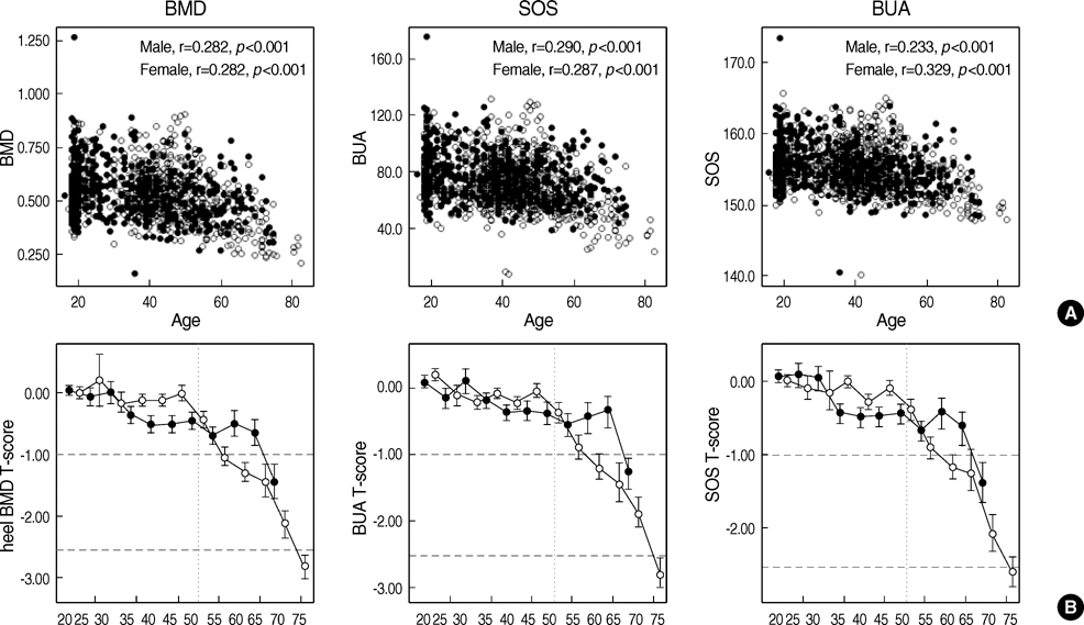
Fig. 3 Age-related changes in QUS data. (A) Actual raw data of QUS parameters with respective correlation values. (B) T-score of QUS parameters. Filled circle, male; void circle, female.
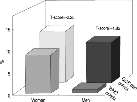
Fig. 4 Comparisons of the prevalence of osteoporosis in Korean women and men, according to either the classical WHO T-score of under -2.5 or the newly-derived heel BMD T-score from QUS adapted from the actual reported occurrence of hip fracture (noted above the each bar)
Reference
-
1. World Health Organization. Assessment of Fracture Risk and Its Application to Screening for Postmenopausal Osteoporosis: report of a WHO study group. 1994 WHO Technical Report Series. Geneva:2. Cummings SR, Kelsey JL, Nevitt MC, O'Dowd KJ. Epidemiology of osteoporosis and osteoporotic fractures. Epidemiol Rev. 1985. 7:178–208.
Article3. Kaufman JJ, Einhorn TA. Ultrasound assessment of bone. J Bone Miner Res. 1993. 8:517–525.4. Langton CM, Palmer SB, Porter RW. The measurement of broadband ultrasonic attenuation in cancellous bone. Eng Med. 1984. 13:89–91.
Article5. Langton CM, Riggs CM, Evans GP. Pathway of ultrasound waves in the equine third metacarpal bone. J Biomed Eng. 1991. 13:113–118.
Article6. Siris ES, Miller PD, Barrett-Connor E, Faulkner KG, Wehren LE, Abbott TA, Berger ML, Santora AC, Sherwood LM. Identification and fracture outcomes of undiagnosed low bone mineral density in postmenopausal women: results from the National Osteoporosis Risk Assessment. JAMA. 2001. 286:2815–2822.
Article7. Grampp S, Genant HK, Mathur A, Lang P, Jergas M, Takada M, Glüer CC, Lu Y, Chavez M. Comparisons of noninvasive bone mineral measurements in assessing age-related loss, fracture discrimination, and diagnostic classification. J Bone Miner Res. 1997. 12:697–711.
Article8. Kanis JA. WHO Study Group. Assessment of fracture risk and its application to screening for postmenopausal osteoporosis: synopsis of a WHO report. Osteoporos Int. 1994. 4:368–381.
Article9. Kastelan D, Kujundzic-Tiljak M, Kraljevic I, Kardum I, Giljevic Z, Korsic M. Calcaneus ultrasound in males: normative data in the Croatian population (ECUM study). J Endocrinol Invest. 2006. 29:221–225.
Article10. Frost ML, Blake GM, Fogelman I. Can the WHO criteria for diagnosing osteoporosis be applied to calcaneal quantitative ultrasound? Osteoporos Int. 2000. 11:321–330.
Article11. Shin CS. Study of an osteoporosis management program in Korea. Management center for health promotion of Korea. 2007.12. Edelstein SL, Barrett-Connor E. Relation between body size and bone mineral density in elderly men and women. Am J Epidemiol. 1993. 138:160–169.
Article13. Ravn P, Cizza G, Bjarnason NH, Thompson D, Daley M, Wasnich RD, McClung M, Hosking D, Yates AJ, Christiansen C. Early Postmenopausal Intervention Cohort (EPIC) study group. Low body mass index is an important risk factor for low bone mass and increased bone loss in early postmenopausal women. J Bone Miner Res. 1999. 14:1622–1627.
Article14. Ito M, Yamada M, Hayashi K, Ohki M, Uetani M, Nakamura T. Relation of early menarche to high bone mineral density. Calcif Tissue Int. 1995. 57:11–14.
Article15. Melton LJ 3rd, Chrischilles EA, Cooper C, Lane AW, Riggs BL. Perspective. How many women have osteoporosis? J Bone Miner Res. 1992. 7:1005–1010.
Article
- Full Text Links
-
- Actions
-
Cited
- CITED
-
- Close
- Share
-
- Similar articles
-
- The diagnosis of osteoporosis
- Prevalence and Factors associated with Osteoporosis using Quantitative Ultrasound Measurements in Women Farmers
- Assessment of Osteoporosis Based on Changes in SNR and ADC Values on MR Diffusion Weighted Images
- The Assessment of Bone Mineral Density in Postmenopausal and Senile Osteoporosis Using Quantitative Computed Tomography
- Assesment of Bone Strength Using a New Quantitative Ultrasound Device in Chidren with Renal Diseases
