J Korean Med Sci.
2010 Mar;25(3):454-457. 10.3346/jkms.2010.25.3.454.
Anatomical and Electrophysiological Myotomes Corresponding to the Flexor Carpi Ulnaris Muscle
- Affiliations
-
- 1Department of Physical Medicine and Rehabilitation, Korea University College of Medicine, Seoul, Korea. hkkwon@korea.ac.kr
- KMID: 1778043
- DOI: http://doi.org/10.3346/jkms.2010.25.3.454
Abstract
- This study was designed to investigate the incidence of lateral root of the ulnar nerve through cadaveric dissection and to analyze its impact on myotomes corresponding to the flexor carpi ulnaris (FCU) assessed by electrodiagnostic study. Dissection of the brachial plexus (BP) was performed in 38 arms from 19 cadavers, and the connecting branches between the lateral cord and medial cord (or between lateral cord and ulnar nerve) were investigated. We also reviewed electrodiagnostic reports from January 2006 to May 2008 and selected 106 cases of single-level radiculopathy at C6, C7, and C8. The proportion of abnormal needle electromyographic findings in the FCU was analyzed in these patients. In the cadaver study, branches from the lateral cord to the ulnar nerve or to the medial cord were observed in 5 (13.1%) of 38 arms. The incidences of abnormal electromyographic findings in the FCU were 46.2% (36/78) in C7 radiculopathy, 76.5% (13/17) in C8 radiculopathy and 0% (0/11) in C6 radiculopathy. In conclusion, the lateral root of the ulnar nerve is not an uncommon anatomical variation of the BP and the FCU commonly has the C7 myotome. Needle EMG of the FCU may provide more information for the electrodiagnosis of cervical radiculopathy and brachial plexopathy.
Keyword
MeSH Terms
Figure
-
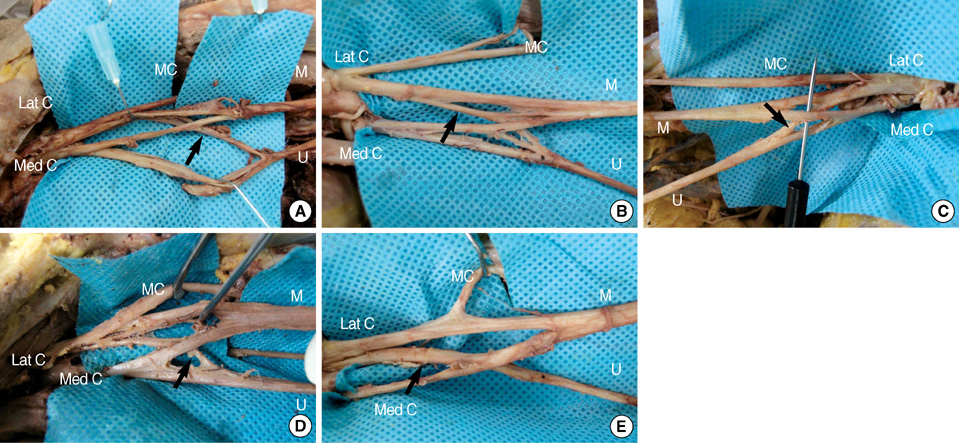
Fig. 1 Five cases of anatomical variations observed in cadaver dissection of 38 upper extremities. Four cases (A-D) show connecting branches (arrow) from the lateral cord to the ulnar nerve (lateral root of the ulnar nerve), and one case (E) shows a connection (arrow) between the lateral cord and the medial cord. Lat C, lateral cord; M, median nerve; MC, musculocutaneous nerve; Med C, medial cord; U, ulnar nerve.
Reference
-
1. Standring S. Gray's Anatomy: the anatomical basis of clinical practice. 2005. 39th ed. Edinburg: Elsevier Churchil Livingston;846–849.2. Perotto AO. Anatomical guide for the electromyographer. 1994. 3rd ed. Springfield: Charles C Thomas Publisher;54–55.3. Aminoff MJ. Electromyography in clinical practice. 1998. 3rd ed. New York: Churchill Livingstone;249–281.4. Haymaker W, Woodhall B. Peripheral nerve injuries: principles of diagnosis. 1998. 2nd ed. New York: Thieme;17–37.5. Dumitru D, Zwarts M. Dumitru D, Amato A, Zwarts M, editors. Brachial plexopathies and proximal mononeuropathies. Electrodiagnostic Medicine. 2002. 2nd ed. Philladelphia: Hanley & Belfus, INC;777–836.
Article6. Kimura J. Electrodiagnosis in disease of nerve and muscle: principles and practice. 2001. 3rd ed. Oxford: Oxford University Press;16–17.7. Lee HJ, DeLisa JA. Manual of nerve conduction study and surface anatomy for needle electromyography. 2005. 4th ed. Philadelphia: Lippincott Williams & Wilkins;172–173.8. Kerr AT. The brachial plexus of nerves in man, the variations in its formation and branches. Am J Anatomy. 1918. 23:285–395.
Article9. Fuss FK. The lateral root of the ulnar nerve. Acta Anat (Basel). 1989. 134:199–205.10. Bowden R, Abdullah S, Gooding MR. Anatomy of the cervical spine, membranes, spinal cord, nerve roots and brachial plexus. 1967. 1st ed. London: Heinemann.11. Ferrante MA, Wilbourn AJ. Electrodiagnostic approach to the patient with suspected brachial plexopathy. Neurol Clin. 2002. 20:423–450.
Article12. Wilbourn AJ. Dyck PJ, Thomas PK, editors. Brachial plexus lesions. Peripheral neuropathy. 2005. Vol 2:4th ed. Philadelphia: Elsevier Saunders;1339–1373.
Article13. Levin KH, Maggiano HJ, Wilbourn AJ. Cervical radiculopathies: comparison of surgical and EMG localization of single-root lesions. Neurology. 1996. 46:1022–1025.
Article14. Leonard JA. Radiculopathy. 1993 AAEM Course D: Fundamentals of Electrodiagnostic Medicine. 1993. Rochester: AAEM;29–36.15. Dillingham TR, Lauder TD, Andary M, Kumar S, Pezzin LE, Stephens RT, Shannon S. Identification of cervical radiculopathies: optimizing the electromyographic screen. Am J Phys Med Rehabil. 2001. 80:84–91.16. Millesi H. Brachial plexus injuries. Management and results. Clin Plast Surg. 1984. 11:115–120.17. Matejcik V. Variations of nerve roots of the brachial plexus. Bratisl Lek Listy. 2005. 106:34–36.
- Full Text Links
-
- Actions
-
Cited
- CITED
-
- Close
- Share
-
- Similar articles
-
- Tendon Problems of the Ulnar Wrist
- Ulnar nerve Compression Syndrome due to anomalous Branch of the Ulnar Nerve Piercing the Flexor Carpi Ulnaris: Report of one case
- Surgical Treatment of Chronic Flexor Carpi Ulnaris Tendinopathy
- Anatomical Relation of Ulnar Nerve and Flexor Carpi Ulnaris Muscle at the Wrist
- Surgical Management of Pisiform Bone Deformity Associated with Tendonitis of Flexor Carpi Ulnaris
